
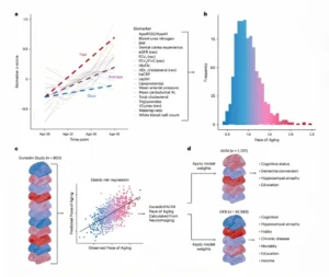
Can a brain MRI reveal how quickly our brain and body are aging? Doctoral student Marta Arbizu Gómez explains how the DunedinPACNI biomarker estimates the pace of aging from a single MRI to predict cognitive decline.
Biological aging does not progress at the same pace in everyone. This article examines DunedinPACNI, an innovative biomarker that can estimate the pace of aging throughout the body from a single brain MRI. Unlike static brain age models, this new longitudinal measure can predict future risk of cognitive decline, hippocampal atrophy, and dementia. Discover the clinical implications of this finding for early assessment, prevention, and the future of personalized neurorehabilitation.
Why do we need to measure the pace of aging?
Aging does not happen the same way in everyone. Two people of the same chronological age can follow very different trajectories: one may maintain good physical and cognitive health for decades, while the other may show early signs of frailty, cognitive decline, or increased disease risk.
For this reason, in recent years there has been growing interest in developing biomarkers that can estimate biological age or, more precisely, the pace at which a person is aging. These biomarkers could help identify people at greater risk of decline early, evaluate preventive interventions, and improve our understanding of how aging relates to conditions such as dementia, stroke, and cardiovascular disease.
Until now, many of these tools have been based on molecular data, such as so-called epigenetic clocks. However, not all studies have access to DNA samples or methylation analyses. Brain MRI, on the other hand, is widely used in neurological research and aging studies.
The study published in Nature Aging raises a very interesting question: Can a single brain MRI provide information about the pace of aging throughout the body?
To answer this question, the authors developed a new measure called DunedinPACNI (Dunedin Pace of Aging Calculated from NeuroImaging), a neuroimaging-based biomarker that estimates the pace of aging from a single brain image.

Subscribe
to our
Newsletter
How was the research conducted?
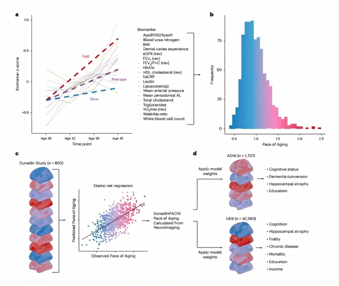
The starting point was the Dunedin Study, a longitudinal cohort from New Zealand that has followed more than a thousand people born between 1972 and 1973 for decades. This cohort is especially valuable because it makes it possible to observe how the health of the same people changes over time, rather than simply comparing people of different ages.
In previous studies, the researchers had calculated the so-called Pace of Aging, a measure of the rate of aging based on 19 biomarkers from different body systems. These biomarkers included cardiovascular, metabolic, renal, immune, pulmonary, and dental indicators, measured repeatedly between ages 26 and 45.
In this new study, the authors used brain MRIs taken at age 45 from 860 participants in the Dunedin Study. From these images, they extracted structural brain measures, including cortical thickness, gray matter volume, cortical surface area, subcortical structure volume, and ventricle size.
Using these data, they trained a statistical model to estimate the Pace of Aging from brain MRI. The result was DunedinPACNI: a measure designed not simply to indicate whether the brain “looks older,” but to estimate whether a person is aging faster or slower in biological terms.
What do the key findings reveal?
The findings show that DunedinPACNI is associated with multiple indicators of brain and overall health.
First, within the Dunedin Study itself, people with higher DunedinPACNI values had a profile consistent with faster aging. They had poorer balance, slower gait, less strength, poorer motor coordination, more physical limitations, poorer self-rated health, lower cognitive performance, and an older facial appearance.
This suggests that information contained in a brain MRI may reflect, at least in part, aging processes that affect the entire body.
The authors also tested the measure’s reliability using independent data from the Human Connectome Project. DunedinPACNI showed excellent test-retest reliability, meaning that the measure remains stable when calculated from repeated images of the same person.
When the researchers applied DunedinPACNI to large external cohorts, such as ADNI and UK Biobank, they found clear associations with cognitive functioning. People with higher DunedinPACNI values performed worse on tests of memory, psychomotor speed, and executive function, and on cognitively demanding activities of daily living.
One of the most important findings was its relationship with cognitive decline and dementia. In ADNI, people with mild cognitive impairment had higher DunedinPACNI values than cognitively healthy people, and people with dementia had even higher values. In addition, among cognitively normal participants, a higher DunedinPACNI value at baseline was associated with a greater risk of developing mild cognitive impairment or dementia in subsequent years.
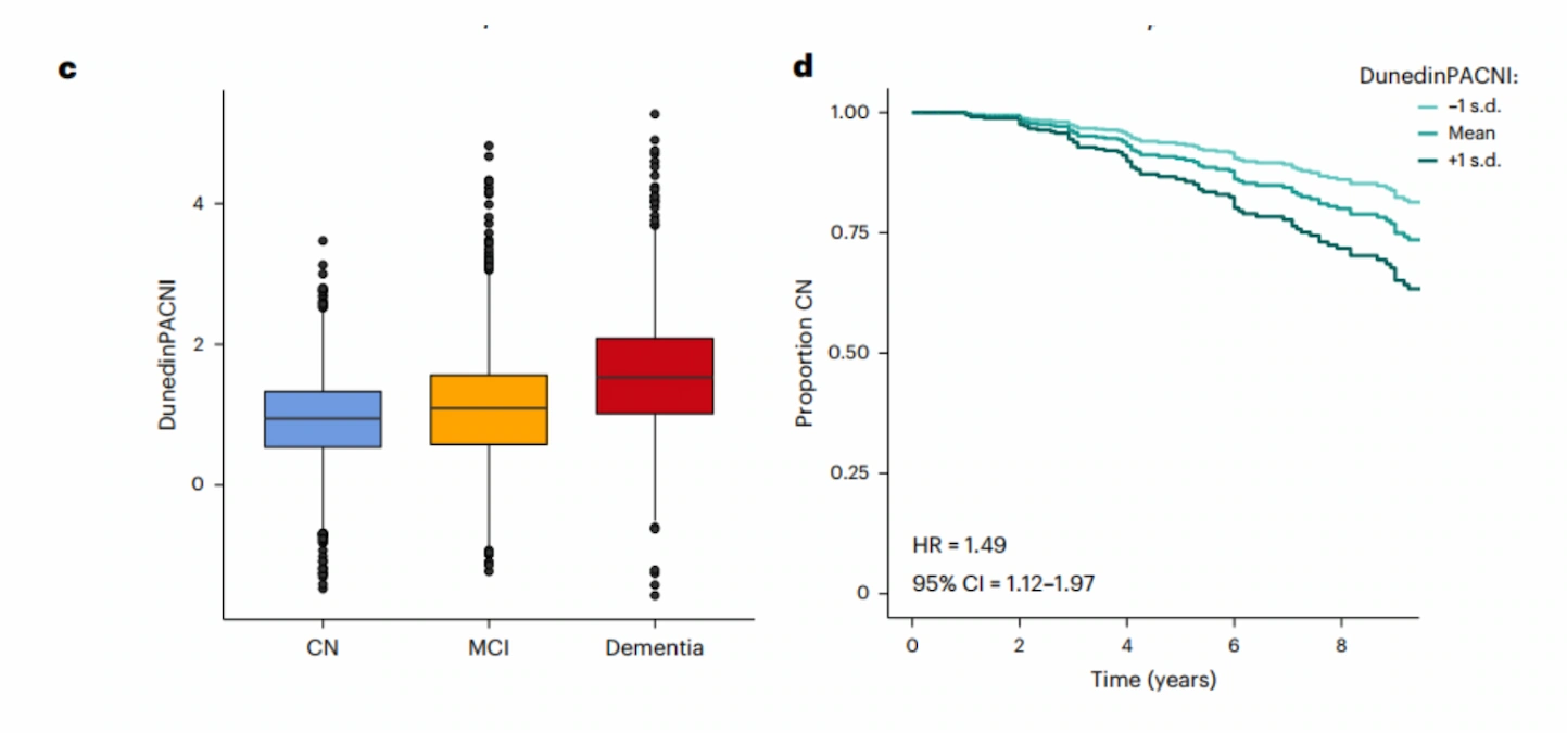
The study also found that DunedinPACNI predicted greater longitudinal hippocampal atrophy. This finding is especially important because the hippocampus is a key structure for memory and one of the regions most affected in Alzheimer’s disease.
But the study is not limited to the brain. In UK Biobank, DunedinPACNI was also associated with greater frailty, poorer self-rated health, a higher burden of chronic disease, greater future disease risk, and higher mortality. In other words, this neuroimaging-based measure appears to capture information related to aging throughout the body, not just brain aging.
| Area assessed | Main finding |
|---|---|
| Validation in the Dunedin Study | DunedinPACNI was associated with poorer physical functioning, poorer cognition, and greater facial aging. |
| Reliability | Excellent test-retest reliability. |
| Cognition | Higher values were associated with poorer memory, psychomotor speed, and executive function. |
| Cognitive decline | Distinguished among cognitively healthy people, people with mild cognitive impairment, and people with dementia. |
| Conversion to dementia | Predicted a greater risk of progression to cognitive impairment or dementia. |
| Brain atrophy | Was associated with greater longitudinal hippocampal atrophy. |
| Overall health | Was associated with frailty, poorer self-rated health, chronic disease, and mortality. |
| Comparison with brain age | Showed associations similar to or stronger than the brain age gap. |
What does it add compared with brain age?
In recent years, the concept of brain age has become popular. These models estimate how many years old a person’s brain “appears” based on its structure. The difference between estimated brain age and actual chronological age is then calculated. If the brain appears older than expected, this is called a larger brain age gap.
However, this approach has some limitations. Predicting chronological age is not always the same as measuring the actual pace of biological aging. A model may be very good at estimating a person’s age without necessarily capturing the processes that explain why some people age faster than others or develop age-related diseases earlier.
DunedinPACNI takes a different approach. Instead of being trained to predict chronological age, it is trained to estimate a longitudinal measure of the pace of aging calculated from body biomarkers measured over nearly two decades.
This distinction is key. The goal is not to determine how old the brain looks, but whether brain structure contains signals of the pace at which the body is aging.
In the study, DunedinPACNI showed associations that were as strong as or stronger than those of the brain age gap for several clinical outcomes, including cognition, hippocampal atrophy, frailty, chronic disease, and mortality. In addition, the two measures were only modestly correlated, suggesting they capture partially distinct and potentially complementary information.
What are the implications for research and clinical practice?
Although DunedinPACNI is not yet a clinical tool, its findings open up several areas of interest.
First, it could be useful in aging studies to identify people whose aging is progressing at an accelerated pace before serious diseases or noticeable functional decline appear.
Second, it could help evaluate interventions designed to promote healthy aging. If a physical, cognitive, pharmacological, or lifestyle intervention aims to slow aging, having an objective measure based on brain MRI could provide additional information about its impact.
Third, DunedinPACNI could complement biomarkers for specific diseases. For example, in Alzheimer’s disease, there are biomarkers for amyloid, tau, or neurodegeneration. DunedinPACNI does not replace these markers, but it could provide a broader view of a person’s biological status and overall vulnerability.
It is also noteworthy that the study explored its use in international samples, including BrainLat, a Latin American cohort of healthy people and patients with Alzheimer’s disease or frontotemporal dementia. In this sample, people with dementia also had higher DunedinPACNI values, suggesting that the measure could generalize beyond the cohorts in which it was developed. Even so, the authors themselves note that more studies in diverse populations are needed.
What are the study’s limitations?
Despite its promising findings, DunedinPACNI should be interpreted with caution.
First, it is a research tool. There are not yet any clinical cutoffs for interpreting an individual’s result, nor sufficiently established normative values for diagnostic use.
Second, many of the cohorts used come from high-income countries and are predominantly made up of participants of European ancestry. Although the study includes validation in a Latin American sample, its generalizability to other populations still needs to be investigated.
Third, DunedinPACNI is based on structural T1-weighted MRI measures. Its accuracy may improve in the future by incorporating other neuroimaging modalities, such as functional MRI or measures of white matter connectivity and microstructure.
Finally, DunedinPACNI estimates the pace of aging from a brain image taken at a specific point in time. This does not replace longitudinal follow-up with repeated assessments, especially when studying an individual’s changes over time.
How does this advance relate to NeuronUP?
At NeuronUP, we develop digital tools for cognitive assessment and rehabilitation based on scientific evidence. Studies like this underscore the importance of integrating different sources of information to better understand each person’s cognitive and functional status.
The possibility of combining brain biomarkers, clinical data, and digital cognitive measures paves the way for more personalized care. For example, in the future, tools such as DunedinPACNI could help identify profiles with greater vulnerability, guide follow-up more effectively, and complement cognitive assessment in people at risk of decline.
In this context, cognitive rehabilitation is not viewed as a stand-alone intervention, but as part of a broader approach to healthy aging. Neuroimaging can provide information about the brain’s biological status; digital platforms, in turn, make it possible to work on specific cognitive functions, track performance, and adapt tasks to each user’s needs.
This type of research points toward more preventive, personalized, and multidisciplinary medicine, in which early detection and cognitive intervention can go hand in hand.
Conclusion
DunedinPACNI represents an important advance in measuring biological aging through neuroimaging. Using a single brain MRI, this tool can estimate individual differences in the pace of aging and is associated with important outcomes such as cognitive decline, hippocampal atrophy, frailty, chronic disease, and mortality.
Its main contribution is that it does not simply calculate whether the brain looks older or younger; instead, it aims to capture signals of the body’s longitudinal aging process. Although further validation is needed before clinical use, DunedinPACNI offers a promising way to investigate how the brain and body age together.
In neurorehabilitation and cognitive health, these findings underscore the need to combine biomarkers, cognitive assessment, and personalized intervention to promote healthier aging.
References
- Whitman ET, Elliott ML, Knodt AR, Abraham WC, Anderson TJ, Cutfield NJ, Hogan S, Ireland D, Melzer TR, Ramrakha S, Sugden K, Theodore R, Williams BS, Caspi A, Moffitt TE, Hariri AR. DunedinPACNI estimates the longitudinal Pace of Aging from a single brain image to track health and disease. Nature Aging. 2025;5:1619–1636. doi:10.1038/s43587-025-00897-z.
Frequently asked questions about brain aging
1. What is the DunedinPACNI biomarker?
DunedinPACNI is a neuroimaging-based measure that estimates a person’s pace of biological aging using data extracted from a single brain MRI.
2. What’s the difference between measuring brain age and the pace of aging?
Brain age models estimate how old the brain appears structurally, whereas DunedinPACNI assesses how quickly the entire body is aging. The latter measure was trained using body biomarkers collected over nearly two decades.
3. Can an MRI predict dementia risk?
Research on DunedinPACNI shows that cognitively healthy people with higher values on this biomarker are more likely to develop mild cognitive impairment or dementia in subsequent years.
4. Which brain structures does this model analyze to estimate aging?
The model uses structural measures obtained from MRI, such as gray matter volume, cortical thickness, cortical surface area, subcortical structure volume, and ventricle size.
5. Is DunedinPACNI ready for clinical diagnosis?
For now, it remains a research tool only. There are not yet any normative values or clinical cutoffs that would allow an individual’s result to be interpreted for diagnostic purposes.





 Talent and Growth: Laura de Juan’s Challenge in Building NeuronUP’s Future

Leave a Reply