
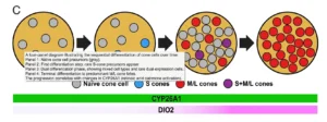
How does the region of the retina responsible for the sharpest vision develop? Doctoral researcher Marta Arbizu Gómez reviews the study that reveals the mechanism.
A recent study reveals the biological mechanism that explains how the foveola forms, demonstrating that photoreceptors change their cell identity during development to build the area of our highest visual acuity. This finding is essential for developing more precise retinal organoids and advancing research into therapies for age-related macular degeneration (AMD). Learn about the anatomical details of this discovery and the importance of neurorehabilitation in mitigating the cognitive impact of vision loss.
What makes the fovea so important to visual perception?
When we read a book, recognize a face, or make out small details, we use a specific region of the retina called the fovea, especially its central area, the foveola. Although it makes up a very small part of the retina, it is responsible for approximately half of our visual perception.
This region has a highly distinctive organization: it is made up almost exclusively of cones sensitive to medium and long wavelengths (M and L cones, responsible for green and red color vision), while it has virtually no blue-sensitive cones (S cones).
For decades, researchers have known that this distribution exists, but it was unclear how it develops during embryogenesis. Now, a study published in PNAS identifies the mechanism that explains how the human retina builds this highly specialized region.
What did the researchers want to find out?
Until now, there were two main hypotheses:
- The first proposed that the different types of cones were already distinct at birth and later migrated to their final positions in the fovea.
- The second suggested that some cones might change identity during development, transforming from one type into another.
To answer this question, the researchers analyzed human retinas at different stages of fetal development and used retinal organoids derived from human stem cells, allowing them to study the mechanisms involved experimentally.

Subscribe
to our
Newsletter
Retinal development: An unexpected change in cell identity
The researchers observed an unexpected phenomenon:
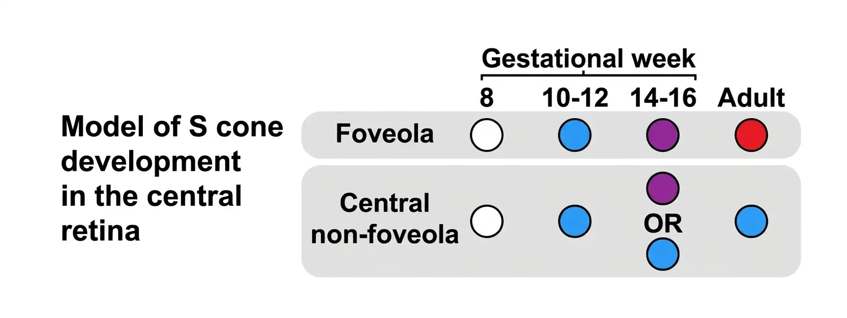
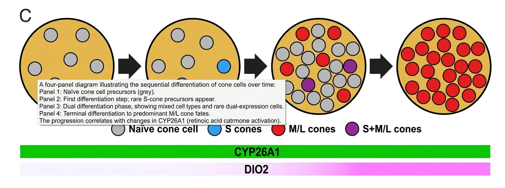
In the early stages of development, some S cones (blue-sensitive) do appear in the future fovea. Over time, however, many of them begin to simultaneously express proteins characteristic of both S cones and M/L cones, until they eventually lose the characteristics of S cones and fully acquire the identity of M/L cones.
In other words, some photoreceptors change identity during development, rather than remaining the same subtype throughout their lives. This gradual change in cone identity during development is summarized in Figure 1.
This finding challenges the traditional view that cell fate is irreversibly fixed once a cell differentiates.
The next step was to identify the biological mechanisms that control this transition.
Key molecular signals in this process
The study also identifies two molecular mechanisms responsible for this organization.
- On the one hand, an enzyme called CYP26A1 reduces retinoic acid levels in the central region of the retina. This decrease promotes the formation of M/L cones and limits the development of S cones.
- On the other hand, sustained signaling by thyroid hormone (T3) drives the few S cones initially present to eventually become M/L cones.
Together, the two mechanisms act in sequence: first, they restrict the generation of S cones, and then they transform those that remain, resulting in the characteristic cellular composition of the foveola.
What do the key findings reveal?
Experiments with organoids made it possible to directly test this mechanism.
When the researchers increased retinoic acid signaling or deleted the CYP26A1 gene, many more S cones appeared and the number of M/L cones decreased.
By contrast, when they maintained prolonged exposure to thyroid hormone T3, the organoids developed a much higher proportion of M/L cones. In addition, some cones initially expressed markers of both types before completing the transition to an M/L identity, confirming that this change in cell identity is part of normal development.
Single-cell RNA sequencing analyses supported these findings and showed that the observed changes did not affect only one specific protein, but the entire gene expression program characteristic of each cone subtype.
Why this discovery matters for macular degeneration research
The foveola is the region most vulnerable to diseases such as age-related macular degeneration (AMD), one of the leading causes of vision loss in older adults.
Understanding how this region forms during development provides valuable information for designing more realistic experimental models and improving future regenerative medicine strategies.
The authors propose that these findings will make it possible to generate organoids with a cellular composition much more like that of the human retina, which could facilitate both the study of disease and the development of cell therapies specifically targeting the macula.
Although this is still basic research, the study provides a mechanistic explanation of how the retinal region responsible for our sharpest vision is organized, and opens up new possibilities for understanding and treating diseases that affect this structure.
The authors summarize this mechanism in a model that integrates the two molecular signals responsible for foveolar development (Figure 2).
How can NeuronUP help?
Visual impairments caused by conditions such as macular degeneration can have a significant impact on independence and everyday cognitive functioning. Although this study focuses on the biological mechanisms of retinal development, improving experimental models may accelerate the development of future therapies designed to preserve central vision. In this context, tools such as NeuronUP can help address functional consequences through tailored cognitive stimulation programs that meet each person’s needs.
References
- Hussey KA, Eldred KC, Guy B, et al. A cell fate specification and transition mechanism for human foveolar cone subtype patterning. Proceedings of the National Academy of Sciences (PNAS). 2026;123(7):e2510799123.
Frequently asked questions about the fovea and macular diseases
1. Why is the foveola so important to visual perception?
The foveola, the central area of the fovea, is responsible for approximately half of our high-acuity visual perception, allowing us to perform complex tasks such as reading or recognizing faces. This region is particularly vulnerable to diseases such as age-related macular degeneration (AMD).
2. How do photoreceptors change identity during human retinal development?
Contrary to the traditional idea of irreversible cell fate, some cones change identity during embryonic development. They initially express S-cone markers (blue-sensitive), transition through a phase in which they coexpress S and M/L markers, and finally acquire a complete M/L-cone identity (red- and green-sensitive).
3. What molecular mechanisms regulate cell specialization in the fovea?
The cellular composition of the foveola is coordinated sequentially by two mechanisms: the enzyme CYP26A1, which reduces retinoic acid levels to promote M/L cones and limit S cones, and sustained thyroid hormone (T3) activity, which transforms the remaining S cones into M/L cones.
4. What are the benefits of stem cell-derived retinal organoids?
Understanding the mechanism of foveal development, as described in studies such as “A cell fate specification and transition mechanism for human foveolar cone subtype patterning.pdf,” makes it possible to generate experimental organoids with a more realistic cellular composition. This supports research into visual disorders and the design of future regenerative cell therapies targeting the macula.
5. How does cognitive stimulation help patients with AMD or central vision loss?
Vision loss secondary to macular diseases directly affects patients’ independence and cognitive performance. Neurorehabilitation tools such as NeuronUP are essential in this clinical context to address functional consequences through personalized cognitive stimulation programs.




 What Do Alzheimer’s and Parkinson’s Have in Common? New Clues From Cellular Transcriptomics
Leave a Reply