

Doctoral researcher Marta Arbizu Gómez addresses an important question: do brain-age models (BrainAge) reflect the specific changes associated with Alzheimer’s disease, or do they capture normal brain aging?
Executive summary:
BrainAge is a biomarker that estimates brain age, but it does not always distinguish between normal aging and Alzheimer’s disease. Studies show that neuroimaging better predicts brain age, whereas neuropsychological tests are more effective for diagnosing the disease. In Spain, combining both approaches can improve clinical assessment and cognitive rehabilitation with tools such as NeuronUP.
What is brain age, and why is it relevant to Alzheimer’s disease?
Aging is the main risk factor for Alzheimer’s disease (AD), but aging and neurodegeneration are not exactly the same. As people age, the brain undergoes natural structural and functional changes. In Alzheimer’s disease, however, these changes accelerate and affect key brain regions involved in memory and cognition.
In recent years, researchers have developed brain-age models (BrainAge), machine-learning algorithms capable of estimating the biological age of the brain from neuroimaging data or cognitive tests. The idea is simple: if a person’s brain appears “older” than their chronological age would suggest, it could be an early sign of neurological decline.
The difference between estimated brain age and actual age is called BrainAge Delta. A positive value indicates that the brain appears more aged than expected, whereas a negative value suggests a “younger” brain.
However, an important question arises: do these models really reflect the changes specific to Alzheimer’s disease, or do they simply capture normal brain aging? A recent study published in NeuroImage and led by the Computational Neuroimaging Laboratory of the Biobizkaia Health Research Institute addresses this question by analyzing how the variables used in BrainAge models influence their ability to identify Alzheimer’s disease.

Subscribe
to our
Newsletter
How was this study on brain age and Alzheimer’s disease conducted?
To answer this question, the researchers used data from the Alzheimer’s Disease Neuroimaging Initiative (ADNI), one of the most widely used international databases in dementia research.
The study included participants from five clinical groups:
- Cognitively normal individuals (CN).
- People with mild cognitive impairment (MCI).
- Patients with Alzheimer’s disease (AD).
- Stable MCI (sMCI).
- MCI that progresses to Alzheimer’s disease (pMCI).
The models were trained using two main types of variables:
Neuroimaging data
Extracted from structural magnetic resonance imaging (T1):
- gray matter volume,
- white matter volume,
- hippocampal volume,
- amygdala,
- thalamus,
- putamen,
- caudate,
- cerebrospinal fluid, among others.
These measures reflect structural changes in the brain associated with both aging and neurodegeneration.
Neuropsychological variables
The researchers also included scores from cognitive tests widely used in clinical practice:
- MMSE,
- ADAS,
- MoCA,
- FAQ,
- ADNI memory score,
- ADNI executive function.
The researchers analyzed which variables were most useful for two different objectives:
- Predicting brain age.
- Distinguishing between different clinical groups.
They subsequently trained different BrainAge models using progressively combined sets of these variables.
Key findings from the study on brain age and Alzheimer’s disease
The results show a clear difference between the types of variables used.
Variables derived from magnetic resonance imaging—such as gray matter or hippocampal volume—were the most informative for predicting brain age. In contrast, neuropsychological tests were more effective at distinguishing healthy individuals from patients with Alzheimer’s disease.
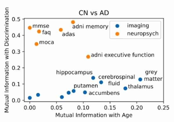
This pattern suggests that models designed to estimate brain age may capture normal aging processes that do not necessarily reflect the pathology specific to Alzheimer’s disease.
Neuroimaging: better for estimating age
Variables derived from magnetic resonance imaging—such as gray matter or hippocampal volume—were the most informative for predicting brain age.
This makes sense because normal aging produces progressive structural changes in the brain.
Cognitive tests: better for identifying the disease
By contrast, neuropsychological tests were more effective at distinguishing healthy individuals, mild cognitive impairment, and Alzheimer’s disease.
In other words, although brain images capture aging well, cognitive performance better reflects the clinical differences between groups.
An important finding: accuracy versus clinical utility
One of the study’s most interesting findings is that there is a trade-off between accuracy in age prediction and the ability to classify disease.
When models are optimized using variables more closely related to aging, age-prediction error decreases. However, their ability to distinguish healthy individuals from patients with Alzheimer’s disease does not always improve. Conversely, when variables with greater discriminatory ability are prioritized, classification of clinical groups improves, although the accuracy of brain-age estimation decreases.
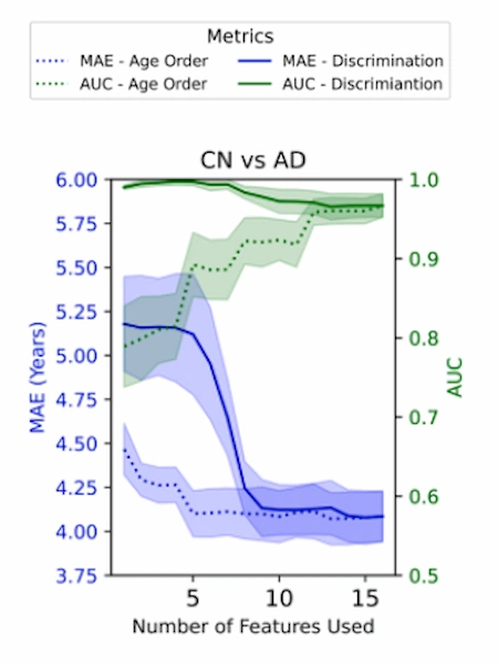
This finding suggests that changes associated with aging and changes associated with Alzheimer’s disease are not exactly the same biological processes.
The key role of the hippocampus
Among all the variables analyzed, the hippocampus stood out as one of the most relevant brain regions.
Its volume showed a strong ability to differentiate between the various clinical groups, consistent with previous evidence: the hippocampus is one of the first structures affected by Alzheimer’s pathology, and its atrophy is closely associated with memory deficits.
Are BrainAge models useful for diagnosing Alzheimer’s disease?
The researchers also compared two different strategies:
- Classifying patients using BrainAge delta.
- Classifying them using the brain variables directly.
The results showed that, in many cases, using the variables directly was equally or even more effective than using BrainAge delta.
This indicates that BrainAge models do not always provide additional information for diagnosis.
However, they offer an important conceptual advantage: they generate a single continuous metric that could be used as a risk indicator or as a tool for monitoring disease progression over time.
What are the implications for Alzheimer’s disease research?
This study provides several relevant conclusions for the development of biomarkers based on artificial intelligence:
- Separating aging from disease is essential. Models that attempt to capture both processes simultaneously may confuse normal aging-related changes with neurodegeneration.
- Variable selection is key. Depending on the objective—predicting age or detecting disease—different types of variables should be used.
- Models should be designed for specific tasks. A model optimized to estimate biological age will not necessarily be the best model for diagnosing Alzheimer’s disease.
Ultimately, aging is not simply the background “noise” in Alzheimer’s disease, but rather a process that actively interacts with the pathology.
How does this advance relate to NeuronUP?
Better understanding the difference between brain aging and neurodegenerative decline has direct implications for cognitive rehabilitation.
On platforms such as NeuronUP, used for cognitive training and the stimulation of executive functions, memory, and attention, reliable biomarkers can help:
- identify patients in the early stages.
- personalize rehabilitation programs according to the cognitive profile.
- monitor patients’ progress over time.
While tools such as BrainAge contribute to improving disease detection and understanding, digital solutions such as NeuronUP make it possible to address patients’ cognitive functioning and quality of life.
This study was led by Dr. Jesus M. Cortes, who is also Director of Research at NeuronUP, from the BioBizkaia Health Research Institute and in collaboration with researchers at Harvard Medical School, University Carlos III of Madrid, Vrije Universiteit Amsterdam, Brigham & Women’s, University of Melbourne, Ikerbasque, and the University of the Basque Country.
Conclusions for professionals
BrainAge models represent a promising tool for studying brain aging and its relationship with Alzheimer’s disease. However, this study demonstrates that the relationship between brain age and neurodegeneration is complex.
Structural brain variables are especially useful for estimating biological age, whereas cognitive tests are more effective at distinguishing between different clinical states.
Therefore, the development of predictive models for Alzheimer’s disease must consider which process is being studied—aging or disease—and carefully select the variables used.
In the future, integrating biological biomarkers, neuroimaging, and digital tools for cognitive assessment and rehabilitation could provide a more comprehensive and personalized view of Alzheimer’s disease treatment.
References
- Garcia Condado J, Verdugo Recuero I, Tellaetxe-Elorriaga I, Birkenbihl C, Carrigan M, Diez I, Buckley RF, Erramuzpe A, Cortes JM. Aging as an active player in Alzheimer’s disease classification: Insights from feature selection in BrainAge models. NeuroImage. 2025. doi:10.1016/j.neuroimage.2025.121548.
Frequently asked questions about brain age (BrainAge) and Alzheimer’s disease
1. What is BrainAge Delta, and how does it help detect accelerated aging?
BrainAge Delta is the metric difference between the brain’s biological age (estimated using AI) and the patient’s chronological age.
- Positive value: Indicates a brain that appears “older” than expected, which is an early warning sign of neurological decline.
- Negative value: Suggests a “younger” brain, which is generally associated with greater cognitive reserve.
In neurorehabilitation centers, this metric makes it possible to objectively monitor the patient’s risk of progression.
2. Is brain age sufficient to diagnose Alzheimer’s disease in clinical practice?
Not on its own. According to the research led by the Biobizkaia Health Research Institute, there is a “trade-off” between accuracy in predicting age and the ability to diagnose the disease.
The models that estimate age best are not always the most effective at distinguishing between a healthy brain and one affected by Alzheimer’s disease. In fact, using brain variables directly may be equally or more effective than calculating BrainAge Delta for clinical diagnosis.
3. Which variables best discriminate Alzheimer’s disease: neuroimaging or cognitive tests?
The study demonstrates that the choice of tool depends on the clinical objective:
- Neuroimaging (T1 magnetic resonance imaging): It is the most informative approach for predicting brain age and capturing natural aging.
- Neuropsychological tests (MMSE, MoCA, ADAS): They are significantly more effective at clinically discriminating among healthy patients, those with mild cognitive impairment (MCI), and those with Alzheimer’s disease.
This confirms that cognitive performance better reflects pathological differences than brain structure alone.
4. Why is the hippocampus key in BrainAge models?
Hippocampal volume stands out as one of the most relevant regions because of its dual role:
- It is essential for estimating biological age.
- It has a strong ability to differentiate clinical groups because it is one of the first structures affected by atrophy in Alzheimer’s disease.
Its analysis is vital for any cognitive stimulation program focused on memory.
6. What implications do these findings have for cognitive rehabilitation?
Understanding the interaction between aging and pathology allows professionals in Spain and Latin America to design more personalized interventions. The use of digital biomarkers and AI models helps to:
- Identify early stages: Detect patients before decline becomes evident.
- Personalize programs: Adjust the stimulation of executive functions and attention on platforms such as NeuronUP according to the user’s biological profile.
- Precise monitoring: Assess the impact of rehabilitation on “brain health” over time.
7. Why is it important to separate aging from disease in Alzheimer’s disease?
Because combining the two processes can lead to misinterpretations. The study demonstrates that BrainAge models may capture normal aging, so it is essential to select variables appropriately according to the clinical objective.





 Chronic Pain Neuroscience: How Pain Reprocessing Therapy (PRT) Retrains the Brain
Leave a Reply