

Doctoral candidate Marta Arbizu Gómez explains how functional connectivity and tumor network mapping are changing the prognosis of diffuse midline glioma.
The prognosis of pediatric diffuse midline glioma (DMG) depends not only on its location, but also on its functional connectivity with healthy brain circuits. Evidence shows that tumors with greater integration into the DMG network are associated with shorter overall survival. For specialists in neurorehabilitation and cognitive assessment, understanding this innovative approach to tumor network mapping is essential for stratifying patients’ neurological risk and designing much more precise, personalized stimulation plans.
Why study brain connections in childhood tumors?
Diffuse midline gliomas, known as DMGs, are pediatric brain tumors that are highly aggressive. They include diffuse pontine gliomas, also called DIPGs, and thalamic gliomas. Despite advances in neuro-oncology, these tumors continue to have a very poor prognosis and show high resistance to the available treatments.
Traditionally, the study of these tumors has focused on their location, molecular characteristics, and radiologic behavior. However, a key idea has emerged in recent years: some gliomas do not grow in isolation but may interact with the brain’s neural networks.
This means the tumor could exploit healthy brain circuits to promote its growth, invasion, and progression. This hypothesis, already observed in preclinical models, raises a highly relevant question: Can the brain connections of the tumor help us predict patients’ clinical course?
The study published in Nature addresses this very question. The authors developed a methodology called tumor network mapping to analyze not only where the tumor is located, but also which brain regions it is functionally connected to.
How was the study conducted?
To study the relationship between tumor, brain connectivity, and survival, the researchers analyzed several independent cohorts of children with pontine or thalamic diffuse midline glioma.
In an initial discovery cohort of 125 patients, the authors segmented tumors on magnetic resonance imaging scans and mapped them to a common brain space. As a first step, they analyzed whether the tumor’s anatomic location alone was sufficient to explain differences in survival between patients.
However, the results showed that tumor location alone did not adequately explain prognosis. The researchers therefore went a step further and studied the functional connectivity of each tumor using pediatric human connectome data. In other words, they calculated which brain regions were functionally connected to each tumor location.
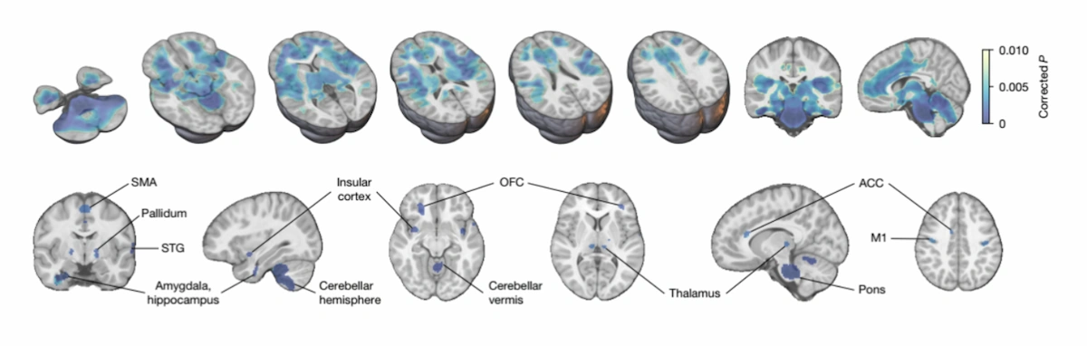
Based on this analysis, they identified a common brain network associated with poorer survival, which they called the DMG network. As shown in Image 1, pontine and thalamic tumors converge on a common brain network. This network includes motor, limbic, thalamic, cerebellar, and brainstem regions, suggesting that the tumor’s functional connectivity can provide prognostic information beyond its anatomic location.
What do the key findings reveal?
The study’s most significant finding is that the tumor’s functional connectivity with the DMG network was associated with patient survival. Specifically, children whose tumors were more strongly connected to this brain network had shorter overall survival.
This finding is important because it changes how we interpret these tumors. Knowing which area of the brain the glioma appears in is not enough; it also seems essential to understand which brain circuits it is connected to. In other words, prognosis depends not only on the tumor’s anatomic location, but also on its integration into a specific brain network.
To confirm the robustness of this finding, the authors validated it in two independent cohorts. In an external multicenter cohort of 125 children, patients with high connectivity between the tumor and the DMG network had a median survival of 8.5 months, compared with 13.3 months for those with low connectivity. The same pattern was observed in a second cohort of 38 children with biopsy-confirmed pontine DMG: greater connectivity with the DMG network was associated with a poorer prognosis.
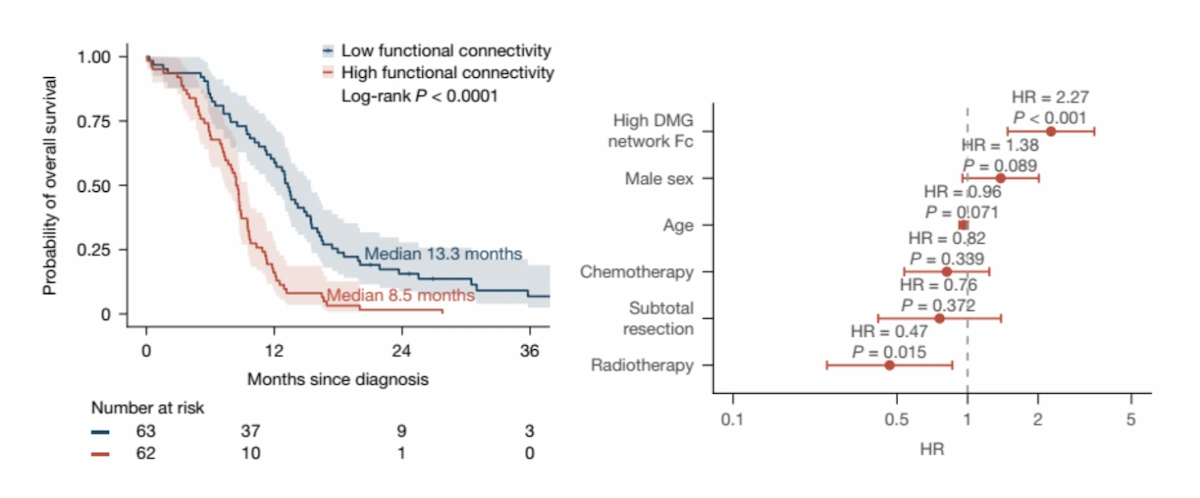
In addition, tumor connectivity provided prognostic information beyond common clinical variables, such as age, sex, surgery, or treatments received. This strengthens the idea that brain network analysis could become a useful tool for better stratifying patient risk.
In summary, the study’s main findings can be summarized as follows:
| Metric / Key finding | Result |
|---|---|
| Discovery cohort | 125 children with pontine DMG/DIPG or thalamic DMG. |
| External validation cohort | 125 children. |
| PNOC cohort | 38 children with biopsy-confirmed pontine DMG. |
| Survival with high DMG connectivity | 8.5 months in the external cohort. |
| Survival with low DMG connectivity | 13.3 months in the external cohort. |
| Main regions of the DMG network | Pons, thalamus, motor cortex, insula, limbic system, cerebellum. |
| Main conclusion | Functional tumor-network connectivity predicts survival better than tumor location alone. |
Another important finding was that tumor growth did not appear to be randomly distributed either. When comparing initial and final MRI scans, the authors observed that tumor progression tended to align with the DMG network. This suggests that tumor spread pathways may be influenced, at least in part, by the brain circuits with which the tumor communicates.
Taken together, the findings point to a central idea: diffuse midline gliomas should not be understood solely as localized lesions, but as tumors that can integrate into functional brain networks. This integration could influence both their progression and patient prognosis.
How is this related to childhood brain development?
One of the most interesting aspects of the study is the relationship between the DMG network and brain development during childhood.
Diffuse midline gliomas do not occur at the same frequency across all ages. Pontine tumors peak in incidence at around 5–6 years of age, while thalamic tumors show another peak at around 10–11 years. The authors analyzed normative data on childhood brain metabolism using FDG-PET and observed that the greatest neurometabolic changes in the DMG network coincided with these ages of peak incidence.
This finding suggests that there are windows of brain development when certain circuits are especially active or vulnerable. In this context, tumors may find a more favorable environment in which to arise, grow, or integrate into neuronal activity.

Subscribe
to our
Newsletter
What do the molecular data show?
The study also analyzed tumor samples using single-nucleus RNA sequencing. Tumors with high connectivity to the DMG network showed enrichment of genes related to synaptic communication and neuron-glioma interactions.
This finding is important because it connects neuroimaging evidence with the tumor’s molecular biology. This is not merely a radiologic association: the more highly connected tumors also show molecular signals consistent with greater interaction with the nervous system.
The authors found signals related to different neurotransmitter systems, including glutamatergic, cholinergic, serotonergic, and noradrenergic systems. Taken together, these findings support the hypothesis that some DMGs can integrate into neural circuits and leverage that communication to promote their progression.
What are the implications for clinical practice?
Although this is a research study and its findings require prospective validation, the implications are highly relevant.
First, the tumor’s functional connectivity could become a new prognostic biomarker. This could enable better patient stratification, identification of higher-risk profiles, and more personalized clinical follow-up.
Second, the study opens the door to new therapeutic strategies focused not only on destroying tumor cells, but also on disrupting the networks that promote tumor growth. This idea is especially interesting for tumors such as DMGs, for which surgical and pharmacologic options remain limited.
In fact, the authors analyzed a subgroup of children with thalamic DMG who underwent subtotal resection. They observed that patients whose resection included tumor areas highly connected to the DMG network had longer survival than those whose less connected regions were resected. Importantly, this effect did not depend solely on the volume of tumor removed, but on which part of the tumor network had been disrupted.
How does this advance relate to NeuronUP?
At NeuronUP, we work with evidence-based cognitive rehabilitation and stimulation tools. Although this study focuses on pediatric oncology and advanced neuroimaging, its findings reinforce a fundamental idea in clinical neuroscience: the brain should be understood as a network.
Neurological changes do not depend solely on a specific lesion or region, but also on how that lesion affects brain circuits. This network-based approach is key to understanding the course of many neurological conditions and designing more personalized interventions.
In childhood brain tumors, understanding how the tumor relates to brain networks could eventually help predict the clinical course, plan treatments, and better understand the functional impact of the disease.
From a cognitive rehabilitation perspective, these advances help us better understand how a brain lesion or disease can affect not just one specific region, but the overall functioning of cognitive networks.
Conclusion
This study proposes a new perspective on the approach to diffuse midline glioma. Rather than viewing the tumor solely as a localized mass, the authors show that its relationship with brain networks may play a key role in progression and prognosis.
Identifying a DMG brain network associated with poorer survival opens new avenues for patient stratification, the design of prognostic biomarkers, and the development of therapies targeting specific brain circuits.
Although prospective studies are still needed to confirm these findings, this work offers a particularly promising idea: understanding brain tumors through connectivity may help us better understand how they grow, spread, and could be treated more precisely.
References
- Sidpra J, Lind V, Cohen AL, Schaper FLWVJ, Stone TJ, Grabovska Y, et al. A prognostic human brain network for diffuse midline glioma. Nature. 2026. doi:10.1038/s41586-026-10631-3.
Frequently asked questions about diffuse midline glioma and brain networks
1. What is diffuse midline glioma (DMG), and what is its prognosis?
Diffuse midline gliomas, known as DMGs, are highly aggressive pediatric brain tumors that include diffuse pontine gliomas (DIPGs) and thalamic gliomas. Despite medical advances, these tumors have a very poor prognosis and show high resistance to currently available treatments.
2. What does tumor network mapping involve?
Tumor network mapping is a methodology that analyzes not only the tumor’s physical location, but also which brain regions it is functionally connected to. This technique has shown that tumors can exploit healthy brain circuits to promote their invasion and progression.
3. How does functional connectivity affect life expectancy in children with DMG?
Research has shown that a tumor’s functional connectivity predicts survival more accurately than its anatomic location alone. Pediatric patients whose tumors have high connectivity with the “DMG network” have a median survival of 8.5 months, compared with 13.3 months for those with low connectivity.
4. Which brain regions make up the DMG network associated with this tumor?
Pontine and thalamic tumors converge on a common brain network (the DMG network) that includes vital and functional regions such as the pons, thalamus, motor cortex, insula, limbic system, and cerebellum.
5. Why does this childhood brain tumor occur more often at certain ages?
The development of these tumors is linked to specific windows in childhood brain development. The greatest neurometabolic changes in the DMG network coincide with the disease’s peak incidence: around 5–6 years of age for pontine tumors and between 10 and 11 years for thalamic tumors.
6. What are the therapeutic implications of disrupting the tumor network during surgery?
Studies indicate that, in patients with thalamic DMG who undergo subtotal resection, survival is longer when tumor areas highly connected to the DMG network are removed. This benefit depends on which part of the tumor network was disrupted, beyond the total volume of tumor resected.
7. How do these findings affect cognitive rehabilitation?
These advances help neurorehabilitation professionals understand that a lesion or tumor affects the overall functioning of cognitive networks, not just an isolated region. This network-based approach is essential for planning treatments, anticipating the clinical course, and designing more precise, personalized cognitive stimulation interventions.




 rTMS and tDCS in Neurodevelopmental Conditions: New Ways to Enhance Neuroplasticity

Leave a Reply