
PhD candidate Marta Arbizu Gómez analyzes a map of one cubic millimeter of cerebral cortex at nanometer resolution; a milestone in connectomics that is key to professional cognitive rehabilitation.
Researchers have achieved a milestone in connectomics by generating one of the most detailed maps of the human brain to date. The discovery of “ultra-strong” multisynaptic connections and a 2:1 glia-to-neuron ratio is redefining the study of neural circuits. For neurorehabilitation experts, these data provide a foundation for more precise and personalized cognitive stimulation programs.
Why do we need detailed maps of the brain?
Understanding how the human brain works is one of the greatest challenges in neuroscience. Although we know a great deal about the brain’s overall organization thanks to techniques such as magnetic resonance imaging and functional neuroimaging, these tools cannot directly show how neurons connect with one another at the microscopic level.
Cognitive functions—such as memory, attention, and learning—emerge from complex networks of neurons connected by millions of synapses. Until recently, however, reconstructing these networks in human tissue with sufficient detail was extremely difficult.
A study published in Science in 2024 takes an important step in this direction. The researchers reconstructed a fragment of human cerebral cortex measuring approximately one cubic millimeter at nanometer resolution, generating one of the most detailed maps of the human brain to date.
This work represents a key advance in the field of connectomics, whose goal is to map all the neural connections in the brain.
How was this human brain research conducted?
The brain tissue analyzed was obtained during neurosurgery to treat epilepsy. Although the fragment was small—approximately 1 mm³ of temporal cortex—it contained enormous structural complexity.
To study this sample, the researchers used serial electron microscopy, a technique capable of capturing images at nanometer resolution. The tissue was cut into thousands of ultrathin sections and subsequently reconstructed digitally.
In total, the study generated:
- More than 1.4 petabytes of imaging data.
- Approximately 57,000 identified cells.
- Nearly 150 million detected synapses.
These data were analyzed using advanced machine learning and three-dimensional reconstruction tools, which made it possible to identify neurons, glial cells, blood vessels, and synaptic connections within the volume studied.
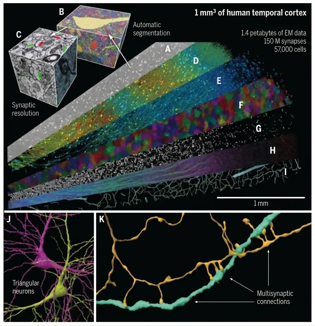
The figure above illustrates the enormous structural complexity contained in an extremely small volume of cerebral cortex, highlighting the technical challenge of reconstructing human neural circuits at this scale.
What does the microscopic structure of the cerebral cortex reveal?
The detailed analysis made it possible to describe how brain tissue is organized at the microscopic level.
In the volume studied, the neuropil—the network of neuronal and glial processes—was composed approximately of:
- Unmyelinated axons: ~40%
- Dendrites: ~26%
- Glial processes: ~15%
- Cell bodies: ~9%
- Myelinated axons: ~7%
In addition, the researchers observed that glial cells outnumber neurons by a ratio close to 2:1, which confirms the fundamental role of these cells in brain function.
The study also made it possible to identify the six layers of the cerebral cortex, each with specific cellular characteristics.

Subscribe
to our
Newsletter
How are connections between neurons organized?
One of the most interesting aspects of the work is the analysis of synaptic connections.
The researchers identified approximately 150 million synapses, classified as:
- Excitatory synapses: ~67%
- Inhibitory synapses: ~33%
They also observed a characteristic organizational pattern:
- Inhibitory synapses are more frequently concentrated near the cell body.
- Excitatory synapses are found primarily on dendritic spines.
This distribution reflects the balance between excitation and inhibition that enables neural circuits to function stably.
New types of neuronal organization
The study also identified structural features that have rarely been described until now.
For example, the researchers analyzed a particular type of neuron in the deep cortical layers called “triangular neurons.” These cells have a large basal dendrite oriented in specific directions.
Surprisingly, the neurons cluster in two main, symmetrical orientations, which suggests a previously unknown structural organization in these cortical layers.
This finding could help improve our understanding of how circuits are organized in deep brain regions.
Surprisingly strong synaptic connections
Another interesting discovery concerns the strength of neuronal connections.
In most cases, an axon forms a single synapse with a target neuron, representing approximately 96% of connections.
However, the researchers found rare cases in which the same axon formed many synapses with the same neuron, with more than 50 connections even occurring between the same neuronal pair.
These findings suggest that cortical circuits combine:
- many weak connections
- a small number of very strong connections
This pattern could be fundamental to the efficient transmission of information in the brain.
What implications does this brain map have for neuroscience?
This study demonstrates that it is possible to reconstruct human neural circuits at synaptic resolution, something that until a few years ago seemed technically unattainable.
In addition, the researchers have made the complete dataset and analysis tools publicly available, allowing other scientists to explore brain tissue at an unprecedented level of detail.
As more maps of this type are generated, it will be possible to:
- Better understand the organization of human brain circuits;
- study how these connections change in neurological diseases;
- and analyze how experience and learning modify neural networks.
Ultimately, this work represents an important step toward a deeper understanding of the microscopic architecture of the human brain.
How does this advance relate to NeuronUP?
At NeuronUP, digital tools for cognitive rehabilitation and stimulation are developed based on scientific evidence.
Research in connectomics provides a fundamental basis for understanding how the neural circuits supporting cognitive functions such as memory, attention, and executive functions are organized.
As we gain a better understanding of these circuits, it will be possible to:
- Design more precise cognitive interventions;
- identify patterns of neuronal deterioration in neurodegenerative diseases;
- and develop more personalized rehabilitation strategies.
Thus, advances in understanding brain architecture complement digital cognitive rehabilitation tools, helping improve the quality of life of people with cognitive impairment.
Conclusion
Reconstructing one cubic millimeter of human cerebral cortex at nanometer resolution constitutes one of the most detailed maps of the brain ever obtained.
Although the volume analyzed is small, it contains enormous structural complexity: tens of thousands of cells and hundreds of millions of connections forming highly organized circuits.
This work marks a milestone in the study of brain connectivity and opens new opportunities for understanding how the microscopic structure of the brain supports our cognitive abilities.
References
- Shapson-Coe A, Januszewski M, Berger DR, et al. A petavoxel fragment of human cerebral cortex reconstructed at nanoscale resolution. Science. 2024; doi:10.1126/science.adk4858.
Frequently asked questions about connectomics and cognitive rehabilitation
1. What is connectomics, and why is it crucial for neurorehabilitation?
Connectomics is the field of neuroscience that seeks to map all the brain’s neural connections. Its importance for neurorehabilitation lies in the fact that it helps explain the physical basis of cognitive functions such as memory and attention, which emerge from complex networks connected by millions of synapses. This knowledge is essential for designing more precise cognitive stimulation strategies based on the brain’s actual architecture.
2. What key discoveries does the new one-cubic-millimeter map of the cerebral cortex offer?
This map, one of the most detailed to date, reveals astonishing structural complexity in a tiny volume. The most disruptive findings include:
- The identification of “ultra-strong” multisynaptic connections, in which a single axon forms more than 50 synapses with the same neuron.
- The observation that glial cells outnumber neurons by a ratio of 2:1.
- The discovery of “triangular neurons” in the deep layers with a previously unknown symmetrical organization.
3. How many cells and synapses were identified in this brain fragment?
Despite its small size (approximately one cubic millimeter), the researchers digitally reconstructed:
- Nearly 57,000 identified cells.
- Approximately 150 million synapses.
This process required generating more than 1.4 petabytes of imaging data using high-resolution electron microscopy.
4. How are excitatory and inhibitory synapses distributed in the cortex?
The study classified synapses into two main types, reflecting the balance required for stable brain function:
- Excitatory synapses (~67%): Located primarily on dendritic spines.
- Inhibitory synapses (~33%): More frequently concentrated near the neuron’s cell body (soma).
5. How does NeuronUP integrate these advances into its cognitive stimulation platform?
NeuronUP uses the scientific evidence provided by connectomics to support its digital rehabilitation tools. By better understanding how the circuits supporting executive functions and memory are organized, it is possible to develop more personalized and effective intervention strategies for people with cognitive impairment or neurodegenerative diseases.

 What Advances in Neuroscience Are Changing Neurorehabilitation in 2026 and How to Apply Them in Clinical Practice
Leave a Reply