

On the occasion of International Epilepsy Day, Psychology student Cristian Francisco Liebanas Vega tells us in this article about the Clinical neuropsychology in pediatric epilepsy neurosurgery. Specifically, he explains what epilepsy is, the types and causes of epilepsy, the stages of the epileptic seizure and the epileptic syndromes that are associated with cognitive manifestations. In addition, Liebanas emphasizes the importance of neuropsychological assessment and neurorehabilitation in patients with epilepsy
Definition and clinical considerations of epilepsy
Epilepsy, like other neurodevelopmental disorders, is not a single pathological entity, and although the hallmark of epilepsy is recurrent seizures, in a significant proportion of children and adolescents it is associated with intellectual problems of cognition and behavior.
Seizures, epilepsy and epileptic syndrome
The common denominator of epilepsy is seizures. According to the International League Against Epilepsy (as of ILAE) a seizure is defined as “the transient occurrence of signs or symptoms due to excessive neuronal activity or synchronous activity in the brain”.
Types of epileptic seizures
Two basic types of epileptic seizures are distinguished:
1. Focal seizure
Localized excitability of cortical or subcortical origin that occurs within networks limited to one hemisphere, and it can be more localized or distributed.
Forms of manifestation
In this type of seizure consciousness may be preserved or altered and different forms of presentation may be observed:
- Onset with motor signs: may present motor automatisms. Different types of seizures can be distinguished, such as:
- Atonic seizures,
- clonic,
- hyperkinetic,
- myoclonic,
- tonic,
- Non-motor onset (automatisms).
- They may present manifestations of automatic function such as sweating, temperature changes, excessive salivation, etc.
- Sensory: tingling, sensations of heat or cold, strong odors, visual disturbances, or pain.
- Cognitive: difficulty with language or a specific cognitive function (aphasia, apraxia or neglect)
- Memory lapses, feeling of Déjà Vu, repetitive thoughts, hallucinations, among others.
- Emotional: intense reactions unrelated to the situation, fear, aggressiveness, crying, abrupt changes (laughing-crying), etc.
2. Generalized seizure
Diffuse across the cortex. They originate at some point within neural networks distributed bilaterally and spread rapidly. These bilateral networks may include cortical and subcortical structures and be asymmetric (Berg et al., 2017).
They can present as:
- Motor: tonic-clonic (tonic and clonic), myoclonic, myoclonic-tonic-clonic, myoclonic-atonic,
- Non-motor: (absences): typical or atypical, myoclonic absence or with eyelid myoclonias.
When a seizure lasts longer than 60 minutes and does not remit with Medication, it is considered refractory status epilepticus. According to the ILAE (2017) this is a condition that can have long-term consequences, including neuronal damage or death and alteration of neuronal networks, depending on the type and duration of the convulsions.
Not all seizures are epileptic; there are many circumstances that can cause acute non-epileptic seizures. It is as important to recognize epileptic semiology as non-epileptic semiology. These can be non-epileptic paroxysmal disorders defined as cerebral dysfunction due to mechanisms different from epileptic seizures. These mechanisms can be:
- anoxic (breath-holding spasm),
- hypnic (night terror),
- psychogenic (anxiety attacks),
- stereotyped movements,
- pathological muscle contractions,
- muscle spasms,
- etc.
Epilepsy is defined as a brain disorder characterized by a persistent predisposition to generate epileptic seizures that has neurobiological, cognitive, psychological and social consequences.
Conceptually, epilepsy exists when the patient has had at least one unprovoked seizure and there is a high probability that a new seizure will occur. In this sense, the ILAE proposed in 2014 an operational clinical (practical) definition of epilepsy which is the one currently in use.
Stages of an epileptic seizure
Every epileptic event usually has 3 stages that must be known and recognized: pre-seizure, seizure onset and post-seizure.
In the pre-seizure phase the precipitating or facilitating factors of the seizure appear: fever, overstimulation, lack of sleep, and so on. It is important to learn which stimuli facilitate seizures in each patient in order to avoid them as far as possible, or at least minimize them.
Sometimes the triggers are environmental stimuli, such as exposure to certain light stimuli or sudden, unexpected sounds. In more severe cases, such as developmental and epileptic encephalopathies, sensory stimulation itself can provoke a seizure. In encephalopathies that involve motor impairment, seizures are sometimes seen to appear when the patient tries to perform a voluntary movement.
At seizure onset the clinical manifestations typical of each type of seizure appear. The most striking are usually generalized tonic-clonic seizures, but there are also far subtler ones, such as eyelid myoclonia.
The post-seizure or postictal phase also has its own clinical manifestations, such as headaches, drowsiness and muscle weakness.
An epileptic syndrome is defined as “a unique cluster of clinical and EEG characteristics, often supported by specific aetiological findings” (structural, genetic, metabolic, immune and infectious).
It frequently carries prognostic or therapeutic implications and often has age-dependent presentations, which means that syndromes generally begin at specific ages and, in some cases, may also remit at certain ages.
Many syndromes are strongly correlated with a range of specific intellectual, psychiatric and other comorbidities, whereas in other syndromes the absence of such comorbidities is a defining feature.
Epileptic syndromes by age of onset
Epileptic syndromes have traditionally been grouped by age of onset. The ILAE describes separately:
- syndromes with onset in neonates and infants (up to 2 years of age);
- syndromes with onset in childhood;
- syndromes that may begin at variable ages (that is, in both paediatric and adult patients).
When epilepsy occurs while the nervous system is still developing, the manifestations typical of neurological disease in childhood appear, and these correspond to a neurodevelopmental disorder.
There are epidemiological, anatomical, logical and pathophysiological reasons to state that epilepsy in children and adolescents is a neurodevelopmental disorder. Above all, there are reasons related to neurodevelopment itself: the immature brain is hyperexcitable, its properties change with each maturational stage, and this determines the appearance of age-specific epileptic syndromes (Mas Salguero, 2020).
Causes of epilepsy
In 2017 the ILAE urged clinicians to classify epilepsy according to its cause and in the context of its comorbidities, such as neurodevelopmental disorders, with the aim of achieving optimal seizure control and improving quality of life. Although the causes of epilepsy in childhood and adolescence are highly varied, three broad groups can be distinguished:
1. Epilepsy caused by a lesion
Caused by any agent that produces permanent damage to the brain: injuries related to prematurity, infections, malformations, trauma, hypoxia, and so on.
2. Epilepsy of genetic origin
Most cases are due to a de novo alteration (Dravet syndrome, for example), and others are clearly hereditary (benign familial neonatal seizures, for example).
3. Seizures that cause epileptogenic lesions
This is the case of acute inflammatory encephalopathies that cause status epilepticus, hemiconvulsion–hemiplegia syndrome (HHS) or febrile infection-related epilepsy syndrome (FIRES), whose cause is unknown.
All of them disrupt the normal functioning of the brain and generate a state of high cerebral excitability which, through different mechanisms, facilitates the appearance of the various types of seizure.
Clinical manifestations by type of seizure
It is extremely important to be able to detect and recognize the most frequent semiology of the different types of seizure. This helps build a clinical pattern of each patient’s epileptic condition, which is very useful when designing the intervention or prevention programme.
Clinical manifestations are sometimes very subtle, which is why it is advisable to ask relatives for a detailed description of the episodes and to record them whenever possible. Depending on where the seizures originate, different clinical manifestations can be observed.
Below are some of the clinical manifestations most frequently seen in paediatric patients.
1. Temporal focal seizures
Patients may present with rising epigastric discomfort (described as burning in the pit of the stomach), increased or new drooling, belching, pallor, flushed face, breath-holding, pupil dilation, fear, panic and olfactory or gustatory hallucinations.
The most frequent symptoms in childhood are automatisms (lip-smacking, chewing or swallowing, facial grimacing, laughter more pronounced on one side of the face, and so on) and a staring gaze with apparent temporary absence.
During the automatism the patient is amnesic, although they may behave as if conscious, if confused. The average duration is 1 to 2 minutes.
Feelings of fear and strangeness are also frequent clinical manifestations in childhood (Etchepareborda, 1999).
2. Frontal focal seizures
- Discharges at the anterior end of the dorsolateral surface of the frontal lobe: these produce generalized tonic-clonic seizures with rapid posterior propagation, abnormal eye movements and loss of consciousness, with the electrical activity spreading quickly towards posterior areas.
- Discharges in the intermediate zone of the dorsolateral surface: these produce complex partial seizures.
- If the electrical discharge is located in the neuronal population immediately anterior to the Rolandic fissure: it produces seizures with motor symptoms that spread following the somatotopic representation of this brain area (starting in a finger and spreading through the whole limb, for example), known as Jacksonian seizures, without altered consciousness.
- If the electrical discharge occurs in the opercular region of the frontal lobe: it produces chewing and swallowing movements, excessive salivation, dysarthria and partial motor signs in the face, without loss of consciousness.
- If the discharge appears in the supplementary motor area, rotation of the head and eyes towards the opposite side is usually observed, accompanied by rotation of the trunk in the same direction, tonic posturing of the contralateral arm and dysarthria (Palacios and Clavijo-Prado, 2016).
- When the electrical discharge affects the cingulate or pericallosal region, the manifestations are: bimanual movements, bipedal movements, brief absences, unmotivated laughter or crying, seizures of terror or rage, and autonomic signs (tachycardia, tachypnoea, pupil dilation) (Etchepareborda, 1999).
- When the discharges appear in the basal or orbital zone of the frontal lobe, gestural automatisms, tonic and clonic movements of all four limbs, olfactory hallucinations, autonomic signs (tachycardia, tachypnoea, mydriasis) and sudden falls appear (Etchepareborda, 1999).
3. Absence seizures
These are a type of generalized epilepsy.
Typical absences involve a disconnection from the environment with an abrupt onset and end and a brief duration. They are sometimes accompanied by motor manifestations, such as oral or ocular movements.
Atypical absences involve a disconnection from the environment with a more gradual onset and end and a longer duration. Patients usually have no memory of the episode.
In some types of absence the patient appears to be conscious and may even produce some speech. There are absences that occur when the patient is engaged in a particular activity, such as reading or writing (Alonso, 2020).
This type of seizure usually affects the patient’s cognitive performance, especially when they occur very frequently: patients return to the task confused and without a goal, which affects how they perform it. They often go unnoticed by family and school and significantly affect learning. They are sometimes misdiagnosed, mainly as ADHD or as a learning disorder.
4. Infantile epileptic spasms
This is an uncommon type of seizure. If it is not treated in time, neurodevelopment may stall or even regress.
Spasms are characterized by head drop, raising and extension of the upper and lower limbs, arching of the back, forward bending at the waist and raising of the knees when lying down. They can also present more subtly, as slight head drops or subtle eye deviations.
It is important not to confuse them with infant colic, an exaggerated Moro reflex or benign sleep myoclonus, among others (Alonso, 2020).
These are some of the clinical manifestations that can be observed in epileptic seizures, but many others may occur that differ in form and frequency from those presented here. It is worth mentioning that in newborns and infants seizures are sometimes subtler or may be confused with non-epileptic paroxysmal events (described earlier); epileptiform pathology should always be ruled out whenever there is any doubt.
Epileptic syndromes associated with cognitive manifestations
Below we briefly describe some epileptic syndromes that present with encephalopathy.
1. West syndrome
An epileptic encephalopathy characterized by infantile spasms, hypsarrhythmia (an EEG pattern) and arrested neurodevelopment involving all areas. There are differences in patients’ neuropsychological patterns: some patients show a pattern with little impairment and others a high degree of impairment. Symptomatic cases, that is, those with a known cause, have a worse prognosis than cases in which the cause is unknown (Fundación Síndrome de West, 2011).
2. Lennox-Gastaut syndrome
The clinical manifestations of this syndrome are tonic seizures predominating during sleep, atonic seizures, atypical absences and progressive neuropsychological deterioration. The later the seizures begin and the fewer the antecedents, the better the response to treatment and the prognosis for cognitive development.
These patients are often refractory to treatment and require pharmacological polytherapy, which further aggravates the cognitive symptoms (Fundación Síndrome de West, 2011).
3. Dravet syndrome
A rare epileptic syndrome characterized by three progressive phases.
Epidemiology of epilepsy and physiology of the developing brain
The incidence of epilepsy by age follows a “U”-shaped curve, with maximum values in the first ten years of life which then stabilize before rising again at later ages.
The incidence of childhood epilepsy is higher at younger ages. It not only interferes with the growth of the brain, delaying the appearance of appropriate adaptive behaviours, but the probability that it appears in the course of a neurodevelopmental disorder is far higher than in the general population, and that is highly significant. The overlap between epileptic syndromes and some neurodevelopmental disorder is very common.
Epileptic encephalopathies appear at early ages and are characterized by an electroencephalogram with abnormal, intractable electrical discharges associated with progressive neurological dysfunction and a considerable impact on neurodevelopment, so that permanent deficits appear in motor skills, cognition and social behaviour that are characteristic of different neurodevelopmental disorders.
Conversely, other conditions that involve a neurodevelopmental disorder (such as Rett syndrome, Fragile X syndrome, trisomy 21 or tuberous sclerosis complex, among many others) carry a high risk of epilepsy, which in turn worsens the prognosis of the underlying condition.
It is no coincidence that the number of people who develop epilepsy is higher at the extremes of life. It is most likely closely related to the brain processes of development in childhood and of degeneration in old age (the formation of connections during growth versus their disconnection in later life).
However, although the synaptic mechanisms disrupted are the same at both ends of life, the results are not the same.
Epilepsy in childhood is more frequent, its types and syndromes more varied, and its pathophysiological mechanisms are intimately linked to the growth of the brain.
The stage of neurodevelopment conditions brain anatomy and physiology and therefore the characteristics of seizures.
Thus, if we look at the incidence of the different types of seizure, we see that myoclonic seizures are the most frequent in newborns, when the primary motor and sensory areas are barely myelinated and epileptic discharges cannot spread. Although far less frequent, focal seizures are also observed at this age.
There are, however, no hemibody or generalized seizures, which can only appear as intracortical connections form and myelination advances.
Following a posterior-to-anterior gradient, the connections of the following regions are successively activated:
- perisylvian – from birth;
- parieto-occipital – between 3 and 7 months of age;
- frontal premotor – from 12 months through the first decade of life.
At the same time, myelination follows a caudal-to-rostral gradient.
All of this contributes to the semiology of seizures in the newborn and the infant, which is determined by these anatomical and functional characteristics as well as by hemispheric specialization and the progressive maturation of the long pathways.
In order to grow, create circuits and modify them, the neurons of the infant nervous system need to be easily excitable, and their synapses need to be facilitatory (excitatory) rather than inhibitory, so as not to impede this development.
We have seen that in the first two years of life the predominant phenomenon of neurodevelopment is synaptogenesis. It happens at dizzying speed (around 1,000 new synapses are formed every second) and these synapses discharge continuously in order to come online and synchronize with one another.
On the one hand, neurotransmission in the immature neuron is fundamentally facilitatory: the GABA receptor promotes the entry of chloride into the cell and there is an abundance of glutamate receptors. Both circumstances favour the passage of the electrical signal by increasing neuronal excitability.
On the other hand, the neuronal circuits being formed have a higher intrinsic resistance (which favours the transmission of action potentials) and emit periodic discharges that help synchronize the new networks, with a discontinuous activity that increases with maturation. This whole excitatory environment facilitates growth and learning, but at the same time favours the development of epileptic mechanisms.
Protective factors against hyperexcitability
Intrinsic protective factors against hyperexcitability
Since epilepsy occurs in only 1–2% of children under two years of age, the brain has intrinsic protective factors against this hyperexcitability:
- a high concentration of neurotrophic factors, which are neuroprotective;
- glutamate reuptake transporters in astrocytes;
- immature cytotoxic cascades that protect against glutamate toxicity;
- a lower concentration of pro-inflammatory cytokines.
Extrinsic protective factors against hyperexcitability
Other, extrinsic factors also act as protectors:
- maternal oxytocin secreted during labour causes chloride to exit the foetal neuron, favouring a more inhibitory action of GABA;
- the newborn’s lipid-rich diet favours the formation of ketone bodies, which have a protective effect on the brain.
From the second year of life, synapse formation slows down and cerebral myelination is more advanced, which contributes to the fall in the incidence of epilepsy from this age onwards.
Early epilepsy occurs when, on top of this natural hyperexcitability of the developing brain, injury mechanisms specific to childhood come into play (genetic defects, defects of neuronal migration, infectious or traumatic inflammatory agents and tumours). These can act at the presynaptic or postsynaptic level, on the glia or on cell adhesion molecules. They also interact with one another, making them much more complex to study.
Alterations in synaptic proteins cause dysfunction of neuronal circuits and play a part in the aetiological and injury mechanisms of both childhood epileptic syndromes and conditions related to autism or intellectual disability.
Genetic studies of childhood epilepsies
Indeed, the genetic study of childhood epilepsies has led to the recognition of genes whose alteration and loss of function has adverse effects on neurodevelopment, with or without epilepsy.
This is the case of the SCN2A gene, which encodes the alpha-2 subunit of the sodium channel. Its mutations can cause disorders of varying severity, including:
- benign familial infantile seizures (which disappear at two years of age with no long-term consequences);
- an infantile epileptic encephalopathy (with seizures that are difficult to control before the first year of life, followed by developmental delay);
- or an autism spectrum disorder in which only 30% of those affected will develop epilepsy, in this case after twelve months of age.
These cases of epilepsies that share a genetic origin with neurodevelopmental disorders have prompted changes in the ILAE classification of the epilepsies, which replaced the term “epileptic encephalopathy” (2010) with “developmental and epileptic encephalopathy” (2017).
This is a further step towards recognizing that childhood and juvenile epilepsy should be considered, without reservation, a neurodevelopmental disorder, and there is a growing body of research supporting this premise.
Neuropsychological assessment and the importance of the clinical neuropsychologist in paediatric epilepsy
Today, great importance is attached not only to the neuropsychological assessment of patients with epilepsy but also to the role of the neuropsychologist within epilepsy teams and units, in diagnosis, surgical intervention and rehabilitation alike.
In the paediatric population, neuropsychological assessment and follow-up provide essential information for the management and guidance of children with epilepsy, since the effect of this disease on a developing brain can lead to the establishment of extensive dysfunctional networks (Fournier, 2019).
The objectives of neuropsychological assessment can be understood from a diagnostic standpoint, since cognitive or behavioural alterations can provide important data for localizing and lateralizing seizures, which is very useful in epilepsy surgery. But it also helps in understanding the epileptic syndrome as a whole, which makes it possible to take better therapeutic decisions, both pharmacological and non-pharmacological.
Objectives of a neuropsychological study in paediatric epilepsy
According to Chelune (2010), the following objectives of a neuropsychological study can be highlighted:
- Establishing a baseline neuropsychological profile in order to identify when there is a risk of developmental, cognitive, academic or psychopathological problems, from the moment the first seizure occurs.
- Helping to detect neurological disorders. In the paediatric population these often go unnoticed and are confused with normal developmental delays.
- Monitoring cognitive and behavioural changes over the course of the disease.
- Diagnosing psychopathological alterations and assessing their impact, both on cognition and on adaptive capacity.
- Helping to make decisions about the planning of therapeutic and educational measures.
- Recording and documenting possible adverse side effects, both cognitive and behavioural, arising from treatment with antiepileptic drugs.
The ILAE recommends that neuropsychological assessment be included in the routine care of patients with epilepsy (Wilson et al., 2015). The role of the clinical neuropsychologist must therefore be an active one and go well beyond administering cognitive tests and analysing them statistically: it requires extensive training and years of experience in the field of both clinical and developmental neuropsychology.
The analysis of cognitive semiology in this type of study is vitally important in order to analyse and synthesize the information provided by the tests administered together with the information gathered from the clinical history and examination of the patient.
Bearing in mind that examining young children with significant neurodevelopmental alterations often makes it difficult to use structured protocols, professionals need to be able to adapt tests and cognitive tasks in order to obtain the cognitive profile that makes the objectives described above possible.
Neuropsychological assessment protocols
Given the characteristics of epilepsy in the paediatric population and its implications for development, it is very difficult to establish standard protocols in the way it is done for adult patients, where several protocols approved by the national and international scientific community are available to guide these assessments.
It is important to understand that we cannot assess cognitive functions or circuits that are still developing or have not yet emerged. It is equally important to consider that in the paediatric population there are more factors that can condition an assessment process and interfere with the patient’s performance during the examination. The main recommendation for paediatric protocols is therefore flexibility, familiarizing the patient with the examiner before starting the protocol, and not treating time as a determining factor.
It is not advisable to use cognitive screening protocols alone: protocols must be broad enough to cover the different cognitive processes and domains that allow us to establish the patient’s neuropsychological profile. We must bear in mind that functions such as memory cannot be reliably explored in children under 5.
When exploring executive functions and certain subcortical processes, we should be aware that the tests available are very sensitive but not very specific, since they can often be altered by antiepileptic drugs which, through their sedative effect, reduce processing speed and attentional maintenance systems. Many tests that depend on information processing will therefore be affected indirectly, preventing us from obtaining objective quantitative data.
It is then that cognitive semiology and qualitative analysis become more important for the conclusions.
Clinical neuropsychology in paediatric epilepsy neurosurgery
Epilepsy neurosurgery in paediatric patients is not usually considered one of the main lines of action. It should not, however, be seen as a last resort for drug-resistant epilepsies.
Given the importance of a developing brain, the main objective of every specialized epilepsy surgery unit team must be, above all, not to worsen the neurodevelopmental prognosis. The contribution of neuropsychology is vital here. The neuropsychologist’s role will be mainly active in the assessment and selection of candidates and at the time of the intervention. Above all, it is key in the post-surgical process, when the neuropsychologist acts as a guide for the family and for the medical and rehabilitation teams alike.
Advances in structural and functional neuroimaging techniques have improved the early detection of potential candidates who present lesions or structural anomalies as the cause of refractory epilepsy (Berg et al., 2017).
Main objectives of a neuropsychological assessment in epilepsy
The main objectives of a neuropsychological assessment, which today forms part of the protocols of epilepsy surgery units according to the ILAE (Jayakar et al., 2014), are:
- to establish a baseline for quantifying the impact of surgery and its results;
- to characterize cognitive strengths and deficits, which are not always detected beforehand;
- to contribute to the localization or lateralization of function;
- to report the risks of deficits after surgery;
- to provide information about children’s educational needs and to plan their rehabilitation.
The standard pre-surgical protocol consists of:
- a high-definition structural neuroimaging study (3 Tesla MRI);
- a neurophysiological study consisting of video-electroencephalography monitoring;
- a complete neuropsychological study.
In order to achieve good post-surgical results (complete resection of the lesion with minimal functional sequelae), it is essential that the epileptogenic zone be located as precisely as possible.
It is therefore essential to distinguish between the symptomatogenic zone — the area of cortex activated by an epileptogenic discharge that produces the ictal symptoms — and the irritative zone, the cortex that generates the interictal discharges, which are the ones we aim to locate with the electroencephalogram (EEG) and magnetic resonance imaging (MRI).
These discharges generally produce no symptoms, since symptoms require very intense after-discharges; most symptoms are generated by propagation. Only occasionally, if the seizure originates in an eloquent area, does it produce clinical manifestations (Rosenow and Lüders, 2001).
Complete resection of the lesion is not always necessary to obtain a good clinical result, and sometimes seizures persist despite complete resection (López and Pomposo-Gaztelu, 2001). This is mainly because structural studies only provide an outline of the lesion, “the tip of the iceberg”, and many of the final decisions about whether to perform a complete resection are therefore taken during the intervention, when direct information about the lesion is available. This is why the neuropsychologist is needed in the operating theatre, even when the patient is not woken on every occasion.
This role is even more important in paediatrics, since the neuropsychologist builds a direct relationship with the child and a bond of trust with the family.
Brain plasticity
The potential for brain plasticity in childhood is critical when planning an intervention, not only because of the capacity to reorganize cognitive functions but also because of the disruption that chronic seizures cause to neurodevelopment, leading to greater cognitive delays.
The neurocognitive development of children undergoing interventions will therefore depend on multiple factors:
- the aetiology of the lesion;
- the age at seizure onset;
- the age at the time of the intervention;
- the type of intervention and any complications;
- pharmacological treatments;
- the context prior to the intervention;
- access to previous neurorehabilitation interventions, and so on.
It is nevertheless clear that if total control or a significant reduction of seizures can be achieved along with the withdrawal or reduction of antiepileptic drugs, patients’ cognitive development will show significant progress.
Most of the information available comes from single cases and experiences or short series. It is necessary to document the neuropsychological progress of these patients so that the risks and benefits of early radical interventions can be better analysed (Fournier, 2019).
The specific protocols are similar to those presented above, with nuances depending on the case: certain functions such as language, memory or visuospatial function can be explored in greater depth.
Psychopathological scales can also be included in order to rule out or confirm alterations of mood or personality.
Patient follow-up
What varies most significantly, however, is patient follow-up. In a surgical protocol it is important to assess the child’s level of consciousness and basic functions on waking from the intervention, as well as in the following days before hospital discharge, and to carry out a complete post-surgical assessment of the neuropsychological profile at 6 months. The main objective is to set the guidelines for the rehabilitation programme and the educational support each case requires.
In some cases, because of the patient’s age (generally 7–8 years) or the type of lesion and its relationship with eloquent areas, an intraoperative assessment with intraoperative brain mapping may be considered. In these particular cases, training the patient in the assessment and control procedures with the referring neuropsychologist should form part of the standard protocol.
Nevertheless, the aim is usually to avoid subjecting a paediatric patient to this type of protocol, first analysing the case with complementary studies such as functional MRI or the Wada test.
When the data obtained are inconclusive or the risk to an eloquent area is very high (especially for language and memory), intraoperative brain mapping is necessary. Where possible (since it depends on each hospital’s resources), it is advisable to run a neuropsychological assessment protocol in the months before the intervention, with the following objectives:
- to assess the patient fully and longitudinally;
- to build a relationship of trust with the child and the family;
- to run a simulated rehearsal of the tests that will be used during brain mapping.
In this period before the intervention it is also advisable to develop a neuropsychological rehabilitation and stimulation programme, which allows us to obtain not only better results in the pre-surgical study but also better results in the operating theatre, and to avoid possible anxiety attacks that could even lead to post-traumatic stress disorder.
Cognitive neurorehabilitation in epilepsy
Factors in patients with epilepsy to consider when designing a neurorehabilitation programme
The neuropsychological pattern in patients with epilepsy varies depending on several factors. The main ones to consider in the neurorehabilitation programme are:
1. Age at seizure onset
As noted above, age is a determining factor in the pathophysiology of the different types of seizure and epileptic syndrome, since the structural and functional changes the brain undergoes from birth to adolescence condition the clinical and neurophysiological expression of the epilepsies.
The moment at which symptoms appear and the previous state of maturation of brain functions define how neurodevelopment is affected.
Early onset of epileptic seizures leads to a greater risk of cognitive impairment. Several studies suggest that the most critical period, with the greatest impact on neurodevelopment, is from 0 to 5 years (Mauri et al., 2001).
2. Types of seizure
Generalized seizures usually involve greater cognitive impairment than focal seizures.
- Generalized seizures affect deep subcortical structures, the thalamus and the brainstem reticular activating system, which are involved in the control of complex brain functions.
- Focal epilepsies are usually associated with specific neuropsychological deficits depending on where the seizures are located (Mulas et al., 2006).
3. Seizure frequency
A higher frequency of epileptic seizures compromises underlying cognitive functioning to a greater degree.
Aetiology of the epilepsy or epileptic syndrome
Epileptic encephalopathy (such as West syndrome or Lennox-Gastaut syndrome, for example) has the worst prognosis, since it involves progressive deterioration of brain function including, over its course, a progressive neuropsychological impairment, probably secondary to abnormal, excessive and diffuse electrical activity (Nieto, 2011).

Subscribe
to our
Newsletter
4. Pharmacological treatment
The effects of antiepileptic drugs can have a positive influence on patients’ cognition and emotion by achieving control of epileptic seizures; however, by also acting on neuronal circuits that regulate cognition, they can produce undesirable neuropsychological effects.
Although currently the new antiepileptic drugs have a lesser effect on cognition, it is still evident in daily practice with these patients that there is a relationship between the drug’s effects and cognitive performance. The problems observed usually refer to specific cognitive deficits rather than a generalized cognitive dysfunction.
Monotherapy
Drug concentration levels in the blood are abnormally high or the rate of dose increase is too rapid (Álvarez- Carriles et al, 2011). Two medications with mild cognitive effects can potentiate each other or produce tolerability problems, leading to cognitive dysfunction (Moog, 2009).
The vast majority of these drugs decrease membrane excitability, increase postsynaptic inhibition or generally alter the synchronization of neural networks and consequently the reduction of neuronal excitability causes a significant decrease in processing speed and of control systems and maintenance of attention, these negative side effects can be as disabling or more disabling than the seizures themselves in developing brains.
General side effects caused by antiepileptic drugs
Although there are significant individual differences in response to treatment, we can establish some general side effects caused by different antiepileptic drugs such as:
- attentional and inhibitory disturbances, aggravated by the drowsiness caused by some antiepileptic drugs or by the insomnia produced by others;
- slowing of processing speed;
- irritability,
- motor restlessness;
- emotional dysregulation;
- impairment of working memory
- effects on the visual field,
- among others (Campos-Castelló and Campos-Soler, 2004).
More severe impairment is usually observed in those patients who have pharmacoresistant seizures, especially if these had an early age of onset, not only because they tend to have a longer chronic period of the disease, but also because they have tried a large number of treatments.
It is necessary to take into account patients’ drug doses and escalation times, since there is a relationship between dose increase and the increase in cognitive symptoms.
5. Brain plasticity
Brain plasticity and functional reorganization in pediatric epilepsy patients provide interesting and highly valuable data for understanding the idea that functions such as language or memory are flexible and modifiable especially during development.
The chronicity of the disease induced by an epileptogenic zone is associated with the recruitment of homologous areas of the contralateral hemisphere or interhemispheric regions that are not traditionally considered eloquent. They allow us during rehabilitation programs to use techniques to optimize or compensate functions—functional alterations that are slowly progressive can change functions such as language from the hemisphere classically understood as dominant (left) to the right, or can redirect language networks during development to non-traditional areas in the same hemisphere (Brazdil et al., 2005). But even so, we must always consider the capacity for functional reorganization as a unique and very complex individual process that requires intervention and follow-up by professionals specialized in the field of neuropsychological rehabilitation.
These are some of the factors that differ in each patient and influence the neuropsychological pattern; nevertheless, in epileptic pathologies there are cognitive functions that tend to be affected to a greater or lesser extent in most cases.
It is common to find involvement of subcortical features, either primarily produced by the abnormal paroxysmal activity or secondarily as a consequence of pharmacological treatment; or the combination of both, which is what we most frequently encounter.
That is, there is a primary subcortical involvement (attention maintenance, working memory, processing speed, Categorical Evocation) that is accentuated and significantly worsened with the use of certain drugs.
Clinical and neuropsychological manifestations observed in patients with epilepsy
Fundamentally the clinical and neuropsychological manifestations observed in patients with epilepsy affect attention, memory, language, processing speed, inhibition and working memory.
In the case of developmental epileptic encephalopathies there is greater overall involvement, both in cognitive and motor processes, so most patients will require interdisciplinary teams in the different contexts of intervention.
The symptomatology present in patients with epilepsy is often the same or similar to that of many neurodevelopmental disorders; therefore, when intervening and setting objectives to stimulate deficits or cognitive alterations, strategies used in other clinical conditions can be used, such as cognitive stimulation, optimization of other cognitive processes or compensation of the lost function.
The most important thing to consider when intervening with a child with epilepsy is the semiology of the seizure, as well as its evolution, since they will determine the cognitive profile and evolution.
Epilepsy occurs in other conditions
It should not be forgotten that epilepsy occurs in other conditions such as cerebral palsy, genetic syndromes, autism, etc., worsening the clinical manifestations of these cases, sometimes being the factor that causes neuropsychological regression.
It should be noted that any type of recurrent seizure affects cognitive functioning, including those labeled as benign childhood seizures. Most patients with benign childhood seizures do not usually manifest symptoms at the onset of seizures, but in the long term subtle symptoms of brain dysfunction begin to appear (mainly related to attention maintenance, working memory and inhibitory control processes). Therefore in these cases it is important to carry out evaluations.
Requirements of a neuropsychological rehabilitation program for epilepsy
Every neuropsychological rehabilitation program in epilepsy must meet the following requirements:
- Be based on theoretical reference models;
- adopt an interdisciplinary and multiple perspective, in the child’s different contexts (therapies, school, family, etc.);
- it is essential to establish an order of priorities;
- important to begin intervention early;
- use a sufficient treatment duration (depending on the case);
- preserved abilities are the basis of treatment;
- very important to consider emotional and motivational variables;
- have good family support.
The importance of neurorehabilitation in patients with epilepsy
Considering all of the above, it should be noted that neurorehabilitation is important not only for cognitive stimulation and rehabilitation, but also for mapping the patient’s cognitive profile and the seizure pattern.
The neuropsychological treatment allows ongoing clinical follow-up of the patient’s evolution, enabling the neuropsychologist to know the semiology presented by each epileptic patient. It also allows identification of different clinical manifestations or changes in the seizure pattern, which may be due to multiple factors (modifications in pharmacological treatment, the patient’s emotional state, overstimulation, etc.).
Follow-up of the patient during neurorehabilitation sessions will allow the neuropsychologist to observe whether there is progressive cognitive deterioration and therefore evidence disease progression, as well as objectify the possible impairment of cognitive functions due to Medication.
This last aspect is important because if we are in contact with the medical team, the neuropsychologist’s observations can contribute to the choice of more effective drug doses that have fewer cognitive adverse effects for the patient.
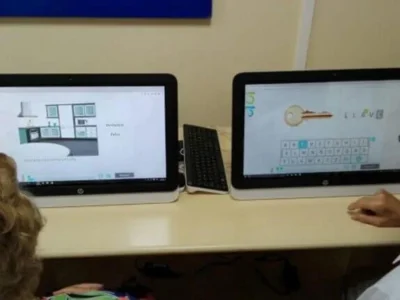
Finally, we want to emphasize that all programs (both computer-based and pencil-and-paper) are tools to work on the affected processes. They do not rehabilitate by mere application; the success of the program depends on the goal we have and the procedure used.
Cognitive processes cannot be understood as independent entities, especially in a network pathology such as epilepsy; the human cognitive system is based on the interaction of different neuropsychological processes, influencing one another both in their development and in their recovery; therefore, we must rule out the rehabilitation of specific functions.
If you liked this post about Clinical neuropsychology in pediatric epilepsy neurosurgery, you may also be interested in the following articles:

“This article has been translated. Link to the original article in Spanish:”
Neuropsicología clínica en la neurocirugía de la epilepsia pediátrica

 A Brief Neuropsychology of Authoritarianism



Leave a Reply