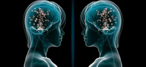
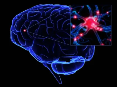
Definition of the mirror neuron system
Neuroanatomy of the mirror neuron motor/imitation system:
There are two main neural networks that make up the mirror neuron system (Cattaneo & Rizzolati, 2008): one formed by areas of the parietal lobe and the premotor cortex, as well as the caudal part of the inferior frontal gyrus; and another formed by the insula and the anterior medial frontal cortex.
We will focus right now on the first system, which involves learning based on observation and imitation. The anatomical organization of the first system follows a somatotopic hierarchy of the ventral premotor cortex, with leg motor acts located dorsally; facial behaviors located ventrally; and manual actions with an intermediate distribution. The location of proximal motor acts (moving the hand toward a point) is represented dorsally, while the simple act of grasping produces ventral activity in the premotor cortex. On the other hand, observation of motor acts also produces differential activation in the parietal cortex.
Observation of transitive acts produces an activation of the intraparietal sulcus, as well as activation of the parietal convexity adjacent to that area. Observation of intransitive acts—whether they are symbolic acts or mime repetition—finds specific activity in the posterior supramarginal gyrus, which extends to the angular gyrus. Finally, observation of acts performed with tools specifically activates the most rostral part of the supramarginal gyrus.
The mirror neuron system produces an evocation of the observed motor actor within the premotor cortex itself. This activity is coordinated simultaneously in the parietal lobe. It is necessary to differentiate the sequence of observation processes to correctly delineate the neuroanatomy of the first exposed system (frontoparietal). In this system, we will speak of observed behaviors that constitute visuomotor priming for the execution—or not—of an action.
Therefore, we will exclude the conception of motor-visual priming, which involves predictions of consequences during action planning. The activity of this frontoparietal system—and here is the relevant point—occurs when the behavior exists—potentially—in the subject’s repertoire. That is, a human observing a bark does not activate premotor and parietal areas, since they do not possess that behavioral repertoire in the cortex.
Moreover, the system’s activity is proportional to the observer’s experience in the behavior being observed. The functional connectivity of the frontoparietal mirror neuron system presents a sequence during observation. Originally this sequence originates in the occipital lobe, where the main features of the observed stimuli are registered. All the information is sent to integration areas in a series of steps that vary from 20 ms to 60 ms, in this order: first the superior temporal sulcus, then the inferior parietal lobe, after that the information goes to the inferior frontal gyrus and, finally, to the primary motor cortex.
Iacoboni et al. (1999) propose that the frontal areas that are activated imply a computation of the goals to be achieved, while parietal activity corresponds to the activation of motor representations of the acts being observed. However, Iacoboni’s group makes a functional differentiation in the neuronal activity of the system, focusing on the pars opercularis of the left inferior frontal gyrus. For them, the dorsal part of the pars is activated when the act is observed and when it is imitated; but ventral activity only occurs when it is imitated. In fact, Iacoboni et al. (2005) functionally analyze the activations described above.
For them, the mirror neuron system is fundamental for learning through imitation. And the activation sequence would be completed as follows:
- (i) first, there is activation of the superior temporal sulcus, where the representations—ventral route of the observed movements—are located.
- From there (ii) it moves to a coding of action goals through the frontoparietal system, in which the dorsal prefrontal cortex would compute the different aspects of the action, such as the goal itself or the meaning, storing this information, sending information to the parietal lobe and correcting computations about space.
This efferent information would be sent from the frontoparietal mirror neuron system, through the pars opercularis, back to the superior temporal sulcus. At this point, a computation of the adjustment between the predicted consequences in the planned imitative action and the visual description of the observed action would occur. In short, the frontoparietal mirror neuron system constitutes a feedback-based learning system.
In fact, what is transferred from visual areas to motor areas is not a detailed motor program, but a prototype of the action, an action with meaning that is processed in the pars opercularis of the inferior frontal gyrus; and which then guides motor planning according to a precise detailed representation of the observed action, represented in the superior temporal sulcus and in the inferior parietal lobe. When the observed action is novel, before the execution period there is activation of the frontoparietal mirror neuron system, in addition to activation of area BA 46 and the anterior medial cortex.
This activation translates into an executive control mechanism, probably as part of Shallice’s supervisory mechanism on which Baddeley (2000) is based to formulate the working memory mechanism. In our case, this system could involve a computation of top-down movement planning, in which working memory handles the observed contents and plans the movement based on them, producing frontoparietal activity that corresponds to the mirror neuron mechanism.
The mirror neuron system should not be conceived as a separate neuronal module, but as an intrinsic and underlying mechanism in most areas related to motor movements. In fact, and as we will see later, disruption of this system does not produce a selective deficit in focal lesions. Rather, the involvement of this system is observed in developmental disorders of the nervous system and in frontal lobe lesions. Let us examine this latter case below.
Dependency and hierarchy
As mentioned, the mirror neuron system overlaps with other systems, and the control system is no exception, since it suppresses spontaneous imitation behaviors. Frontal lesions cause a series of deficits characterized by the emergence of impulsive behaviors generated by external stimuli. Imitative behavior is especially relevant to the mirror neuron system, and may be part of “environmental dependency syndrome”. Normally, the condition arises from a bilateral lesion, although it can also be due to a unilateral lesion, less frequently. Observing the behavior of others can elicit activation of premotor and parietal areas dependent on the mirror neuron system.
In healthy subjects, this activation does not manifest because there is suppression by the frontal lobe. Its deterioration implies a destruction of these mechanisms, transforming potential acts into acts in fact. Echopraxia constitutes the forced and critical imitation of observed behaviors, normally with the presence of perseverations. Although it usually occurs as a disorder associated with damage to the basal ganglia, it also occurs due to frontal deterioration, which produces a disinhibition of the mirror neuron system.
Functionality of the frontoparietal mirror neuron system
Imitation and learning
A fundamental task of learning is imitation, which produces the development of some basic social developmental skills, especially in the acquisition of gestural and postural identification, and allows the development of understanding others’ intentionality.
These neurons fire when the subject performs behaviors related to a goal, but especially when they observe these behaviors in others, discriminating between the different components of the action according to which are more or less relevant from the intentional point of view; even in the absence of objects.
From the above it follows that mirror neurons not only handle contents related to motor or visual patterns, but also abstract ones, both regarding the sensory modality of the contingency (a sound with meaning) and regarding elements of a non-present or abstract nature, which relate, in terms of learning, to intentionality, a reality in which understanding others’ motives plays an important role.
Integrated motor information presents significant procedural characteristics:
- movement processing,
- processing of body parts,
- monitoring of the action aimed at a goal of an external subject,
- etc.
The proximity to frontoparietal systems that support various types of sensorimotor integration suggests that the action coding implemented in the mirror neuron system is tied to some form of sensory integration. Imitation is one of the many forms of this type of integration. In such integration, the observing subject makes comparisons between the information existing in primary areas (visual inputs) and the observed behavior, as explained above.
The imitation behavior literature emphasizes that an essential aspect in this area is the differentiation between various forms of imitation or contagion, and true imitation—that is, adding something new to one’s motor repertoire after observing others performing that action. This differentiation is observed at the neuronal level, distinguishing the interactions between the mirror neuron system and prefrontal and parietal execution-preparation structures during imitation learning, and the interaction between the mirror neuron system and the limbic system during emotional contagion. Probably, as we will discuss later, the mirror neuron system in autism also allows making this distinction, with one of the interaction systems more damaged than the other.
Mirror neurons have individual properties:
They activate during the imitated action, but also during the action being observed even without imitation. They have two levels of congruence:
- strict, in which neurons activate exclusively on actions and observations that are substantially identical;
- and approximate congruence, in which they activate in response to observing an action that is not necessarily identical to the executed action, but achieves the same goal.
Activation thresholds are defined by the logic of the action, not by the object or the distance of the action. From these properties it can be inferred that they handle abstract contents of the observed actions. But what is the degree of abstraction of this coding? High, as shown in experiments with prior “occlusion” conditions, in which neurons activate based on an initial situation of presence or absence, discriminating situations.
There is sensory recognition of auditory actions (sound inputs) in the mirror neuron system. This allows a basis for the understanding of speech and language as a code that is learned—at least in initial phases—through physical and gestural imitation.
Functional hierarchy of the frontoparietal system in motor processing
As mentioned above, there is a functional hierarchy in the mirror neuron system when the subject observes a motor action in order to learn it. The basic levels of motor processing have been extensively studied. However, the mirror neuron system responds to a hierarchy in which movement processing is high-level, producing computations between the consequences of the action and the goals.
To compute such knowledge, the components that present the context of the action must be dissociated: first, the object itself that represents the goal. Existing studies were not conclusive until relatively recently. However, using neuronal suppression techniques, such as magnetic stimulation, it has been possible to dissociate high-level processing from merely kinematic processing. It has been observed that identification of the object-goal is computed in the anterior intraparietal sulcus (Hamilton & Grafton, 2006). Therefore, there is differential processing of objects, even if the action is the same (for example, grasping). On the other hand, this dissociation also implies the analysis of the expected consequences of the action, which presents a higher level of hierarchy than the former.
It is very important to bear in mind that processing goals implies processing the movements necessary to achieve that goal, but these are aspects with a different level of processing, with the processing of the motor program (not its planning) being a lower processing rank. Hamilton & Grafton (2007) demonstrated that there is a lateralization of the mirror neuron system that computes the consequences of action. They found that the consequences of an observed action are processed in the right inferior frontal gyrus and right inferior parietal lobe, as well as in the left postcentral sulcus and the left anterior intraparietal sulcus.
Together they proposed a hierarchical model composed as follows: on the one hand, there is a low-level—cognitive—processing that involves processing the motor pattern. The processing of the motor pattern takes place in a system that involves both visual and motor analysis of the action. The visual would be carried out in lateral occipital areas, while the processing of the kinematic pattern is carried out in the inferior frontal regions.
High-level processing, defined by an analysis of goals, is carried out in a system that involves two areas of the right hemisphere: the intraparietal lobe and the inferior frontal gyrus—to a lesser extent. In this goal processing, object-goals are also processed in a lateralized way in the left inferior parietal cortex. Is there a neuronal hierarchy when observed actions are executed? Yes, and the hierarchical rank differentiates the complexity of actions, that is, when actions are simple, when they are complex—composed of different steps—, as well as when they respond to an intentionality. In this case, the lateralization of neuronal activity does not seem as evident.
There is evidence that planning of simple acts occurs in the motor and premotor cortex, as well as in the left inferior parietal cortex. However, it seems that the right inferior parietal lobe is involved in complex behaviors that require several steps, such as the Tower of London task (Newman et al., 2003). This area seems important for sending feedback about the consequences of the motor act, and together with the cerebellum can compute corrections of movement in space or in planning.
Language
During the task of imitating finger movement, an increase is observed in activity of the rostral posterior parietal cortex and in the inferior frontal gyrus, areas close to Broca’s area, which suggests the involvement of these mirror areas in a phylogenetic-type language acquisition mechanism (Iacoboni & Dapretto, 2006).
This theory has been supported by various indications. First, a left lateralization of the mirror neuron system has been demonstrated. On the other hand, activation of the mirror neuron system in the macaque brain allows extrapolating its areas to ours: the areas in the macaque would coincide with human BA 44, adjacent to Broca’s area. Based on the theory of semantic expression, which proposes that language is learned in a bottom-up process, and the motor theory of speech perception, which proposes that the target of speech analysis is the facial expressions associated with sounds rather than the sounds, it has been found that during speech perception the motor areas of speech are activated, which coincide with the mirror neuron system.
Moreover, it has been found that processing linguistic material produces motor activation, and that the neuronal activity produced by processing linguistic material related to body parts and actions activates the somatotopic zones of the brain related to reading.
Social cognition and mirror neurons
Neuroanatomy of the limbic mirror neuron system
The second mirror system is the emotional one. As we have said above, this system is involved in adopting empathic behaviors, but it does not necessarily operate separately from the first system, although this point will be addressed later. The mirror neuron system is also located in cortical areas that mediate emotional behavior. Observing another’s pain produces activation of the cingulate cortex, the amygdala, and the insula. The insula is especially important in integrating sensory representations, both internal and external. It has an agranular structure and is cytoarchitectonically similar to motor areas.
Therefore, the insula functions as a communication node between the limbic system and the somatotopic cortical activation associated with pain, both one’s own and others’, which constitutes the evolutionary basis of empathy. However, this basis is not unique. The empathy system would be framed as follows:
- First, there must be a node in this system, which is the amygdala, necessary for the emotional activation of subjects.
- Second, the areas of emotional expression and regulation. As we have said, the emotional expression area based on bodily schemas is composed of two structures: first the insula, which as mentioned is the center for integrating interoceptive information. On the other hand we would find the cingulate cortex, which is subdivided as follows: against the classic division between cognitive/dorsal and emotional/rostral processes of the cingulate cortex (Posner et al., 2007); it has recently been shown that there is a division in terms of emotional expression (interoceptive) in the dorsal anterior cingulate cortex, and a regulatory function of emotions in the rostral anterior cingulate cortex (Etkin et al., 2010), which is congruent with an anteroposterior orbitofrontal control system (orbitofrontal-cingulate-amygdalar, mainly).
- Third, the high-level processing node composed by the mirror neuron system. This system is formed by the insula and the anterior medial frontal cortex. In fact, the insula’s mirror neuron system overlaps with the interoceptive emotional expression system. The interaction of this system with emotion varies depending on the complexity of the emotional act.
How does the mirror neuron system work in social cognition?
The mirror neuron system functions in two ways with regard to social cognition:
- First, it is necessary for prediction and the attribution of mental states (theory of mind).
- Second, it activates mechanisms of affective recognition and expressivity. The first prediction system has been explained: the observed acts are computed in a frontoparietal mirror neuron system, together with the consequences.
This system serves as a predictive model that evolves: from simple bottom-up behaviors and processes, the neurological system over the years moves to a top-down regulatory system, in which observed motor schemas are compared with learning accumulated over the years, and aims to establish statistical predictive patterns that minimize error (Kilner et al., 2007). This computation is hierarchical as well, in the sense that the operations performed correspond to a hierarchical distribution of the brain’s theoretical axes. In this hierarchy, the frontal lobe is responsible for computing between the observed behavior and the supposed mental state, and the motor cortex, the parietal cortex and the superior temporal sulcus would be responsible for integrating the visual information and the stored motor schemas.
Next we will focus on the second empathy system, which involves the limbic mirror neuron system (insula, cingulate cortex and frontal lobe).
Mirror neurons and empathy
The role of mirror neurons in empathic behaviors, such as the adoption of facial gestures and postures in interactive imitative behaviors, is fundamental along with emotional adoption (limbic system). As mentioned above, mirror neurons compute movements in terms of execution consequences and goals. This knowledge serves as a basis for social cognition, together with the second emotional integration system. Empathy is not a univocal process. Although there is evidence that observing another’s punishment produces activation in the amygdala, the anterior cingulate cortex and the insula—as well as the thalamus and the cerebellum—(Jackson et al., 2005), the complete process probably depends on a large-scale network, with areas of high processing influencing or eliciting emotional responses.
In fact, this could be the role of mirror neurons in empathy. Empathy is supported by a large-scale neural network composed of the mirror neuron system, the limbic system, and the insula, which functions as a connector node between both systems. Within this network, mirror neurons provide the simulation of expressions and facial gestures observed in others to low-level processing areas, via the insula, which provokes activity in those areas. And, finally, producing an emotional state in the observer of the observed behavior. In this way an alternate system of emotions is provided to the subject, based on simulation, which partly enables social cognition.
This theory is called the “simulation theory” (Gallese & Goldman, 1998; cited in Frith & Frith, 2006), and proposes that in this way we can understand observed emotions through the internal states they provoke in us. Therefore, the most common way of empathizing is literally adopting the other’s posture to simulate it internally. And again, when we try to adopt the posture of the person expressing their emotions, we do so facially, which activates the limbic system.
In short, mirror neurons present a sensorimotor basis of empathy. When talking about this mirror neuron system and its relationship with empathy, it is necessary to make a distinction: understanding and simulating emotions is not the only step for social cognition, since we must take into account the stable personality of the person in order to make predictions.
In this respect, it is interesting to make, again, a distinction: neuronally, is it the same to think about the likely behavior and emotion of a person similar to us as of a different person? It is not. Thinking about someone who is similar to us usually activates areas of the ventral medial prefrontal cortex, especially BA 18, 9, 57 and 10, while thinking about the probable reactions and characteristics of others activates areas of the dorsal prefrontal cortex, BA 9, 45 and 42 (Frith & Frith, 2006).
In fact, there is a medial-lateral functional axis of the brain, in which the more central areas are related to representation of the self and one’s own emotions, while the lateral regions involve a representation of the external world and others. This hypothesis about a medial-lateral axis is based on the fact that medial areas usually present greater connection with limbic centers and proprioceptive sensory information, and therefore are more influenced by data, while lateral areas would be more reflective and dependent on representations about the external world. Amodio & Frith (2006) mention a central node in social cognition processing: the medial frontal cortex (BA 10).
Mirror neuron system and motor rehabilitation
Although the function of the mirror neuron system in motor learning has been explained, it is interesting to note its implication in the formation of a motor memory bank. The strongest evidence comes from the studies of Stefan et al. (2007), in which the authors show how learning a motor sequence through observation enhances the formation of motor memories compared to solo learning. It has been found that learning by observation can mediate long-term neuroplasticity processes in the individual, and that this effect is mediated by the mirror neuron system in the motor cortex.
In a study by Ertelt et al. (2008) two groups of patients with middle cerebral artery stroke and a paretic limb were subjected to two different therapies: one with audiovisual cues, and another without cues. The group that followed the training with audiovisual samples of the exercises showed greater improvement in the paretic limb than the control group. In addition to the above, mirror therapy has been proposed as an alternative that induces changes in plasticity. In it, the patient practices with their healthy limb in front of a parasagittal mirror in which it is visualized. This produces a visual illusion of the paretic limb. Therapy results show cortical plasticity generation.
Mirror neurons and therapy in autism spectrum disorders (Autism and Asperger’s)
Development and dysfunction
There is indirect evidence of mirror neuron activity from the first year of life for the prediction of goals of observed subjects (Falck-Ytter etal., 2006; cited in Iacoboni & Dapretto, 2006). In children under 11 years, these evidences, although less robust than in adults (which is logical if we think that the system is not yet fully mature in connectionist terms), show indices of mirror neuron activation in several parameters (mu rhythm suppression, EEG, near-infrared spectroscopy, functional magnetic resonance) for imitation activities, social competence and empathy. Therefore, although today it is not known with certainty what degree of involvement the mirror neuron system has in social behavior, it is evident that it plays some central role.
Probably one of the keys that allows establishing its importance is its dysfunction in children with autism and other communication disorders. In autism, a deficit of neuronal simulation in modeling observed behaviors has been proposed, which prevents a correct “experimental understanding” of others. This deficit has been neurologically verified in the mirror neuron circuit, in which there are structural abnormalities in subjects with autism spectrum disorders.
This disorder, for example, prevents a correct identification of emotions in facial gestures because there is not adequate activation of the central circuit. However, autistic subjects are able to identify the non-emotional act—although they do not know the purpose for which it is performed—, which suggests a disruption of the limbic mirror neuron circuit more pronounced than the imitation mechanism. This deficit correlates with the severity of the disorder.
Data provided on Asperger confirm a similar but less severe deficit (based on difficulty and a temporal delay in the acquisition of behaviors) that involves imitation, which suggests that imitation may play an important role in therapy with these subjects. Therapy for subjects with autism spectrum disorders may involve the mirror neuron system. There is empirical evidence that at least in part the disorder involves a deficit in imitation and language production, and that the mirror neuron system is implicated in this disorder (Wan et al., 2010).
It has been shown that music therapy favors symptom improvement. Since the sensorimotor system is involved in language processing, and there is also modulation of motor activity during language processing, it seems logical to think that a way to activate the mirror neuron system could improve both symptoms. And music produces activity in the system, which favors its alteration (in a positive sense) and provides neural plasticity, since music is also an act of motor expressivity, and produces activation, among others, of Broca’s area (BA 44). In fact, this type of therapy combined with singing produces beneficial effects in patients with Broca’s aphasia, many of whom are able to say words integrated in a prosody different from normal.
Compared with the act of speaking, singing produces bilateral activation of a frontotemporal network, and part of this network shares neurons with the mirror neuron mechanism. This overlap produces an improvement of audio-motor coordination schemes, a deficit of activation that is observed in people who suffer from this type of communication disorders.
As for imitation, the degree of implementation and effectiveness varies in the same way as the experimental data described above. It seems that children with Asperger show good progress, especially if therapy is initially carried out with subjects close to the affected person, and much more so if it is done with videos of the subject himself. These data have been verified in the suppression of mu waves in the sensorimotor cortex, part of the mirror neuron system.
References
- Cattaneo, L., & Rizzolatti, G. (2009). The mirror neuron system. Archives of Neurology, 66(5), 557-560.
- Ertelt, D., Small, S., Solodkin, A., Dettmers, C., McNamara, A., Binkofski, F., & Buccino, G. (2007). Action observation has a positive impact on rehabilitation of motor deficits after stroke. NeuroImage, 36 Suppl 2, T164-173.
- Frith, C. D., & Frith, U. (2006). The neural basis of mentalizing. Neuron, 50(4), 531-534.
- Grafton, S. T., & Hamilton, A. F. D. C. (2007). Evidence for a distributed hierarchy of action representation in the brain. Human Movement Science, 26(4), 590-616.
- Hickok, G. (2010). The Role of Mirror Neurons in Speech and Language Processing. Brain and language, 112(1), 1.
- Iacoboni, M. (2009). Imitation, empathy, and mirror neurons. Annual Review of Psychology, 60, 653-670.
- Iacoboni, M., & Dapretto, M. (2006). The mirror neuron system and the consequences of its dysfunction. Nature Reviews. Neuroscience, 7(12), 942-951.
- Jackson, P. L., Meltzoff, A. N., & Decety, J. (2005). How do we perceive the pain of others? A window into the neural processes involved in empathy. NeuroImage, 24(3), 771-779.
- Kemmerer, D., & Castillo, J. G. (2010). THE TWO-LEVEL THEORY OF VERB MEANING: AN APPROACH TO INTEGRATING THE SEMANTICS OF ACTION WITH THE MIRROR NEURON SYSTEM. Brain and language, 112(1), 54-76.
- Kilner, J. M., Friston, K. J., & Frith, C. D. (2007). Predictive coding: an account of the mirror neuron system. Cognitive Processing, 8(3), 159-166.
- Le Bel, R. M., Pineda, J. A., & Sharma, A. (2009). Motor-auditory-visual integration: The role of the human mirror neuron system in communication and communication disorders. Journal of Communication Disorders, 42(4), 299-304.
- Oberman, L. M., & Ramachandran, V. S. (2007). The simulating social mind: the role of the mirror neuron system and simulation in the social and communicative deficits of autism spectrum disorders. Psychological Bulletin, 133(2), 310-327.
- Oztop, E., Kawato, M., & Arbib, M. (2006). Mirror neurons and imitation: a computationally guided review. Neural Networks: The Official Journal of the International Neural Network Society, 19(3), 254-271.
- Rizzolatti, G., Fabbri-Destro, M., & Cattaneo, L. (2009). Mirror neurons and their clinical relevance. Nature Clinical Practice. Neurology, 5(1), 24-34.
- Vogt, S., & Thomaschke, R. (2007). From visuo-motor interactions to imitation learning: behavioural and brain imaging studies. Journal of Sports Sciences, 25(5), 497-517.
- Wan, C. Y., Demaine, K., Zipse, L., Norton, A., & Schlaug, G. (2010). From music making to speaking: engaging the mirror neuron system in autism. Brain Research Bulletin, 82(3- 4), 161-168.
“This article has been translated. Link to the original article in Spanish:”
Sistema de Neuronas Espejo: función, disfunción y propuestas de rehabilitación


Leave a Reply