Doctor of Biomedicine Pablo Barrecheguren talks about the term Brainbow, a technique that allows distinguishing neighboring neurons.
In 1906 the Nobel Prize in Physiology or Medicine was shared between Camillo Golgi and Santiago Ramón y Cajal. Largely it was awarded to Golgi for creating a type of stain that marked neurons and their branching, but this had the problem that, because there were so many neurons, it was very difficult to see where each neuron began and ended.
Cajal modified the stain so that far fewer neurons were distinguishable, which allowed seeing each cell individually and discover that in neuronal communication there are gaps.
The Brainbow
Thus, since the beginnings of histology it has been a constant problem to distinguish neurons and their processes within a stain.
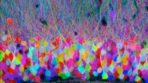
And it’s no wonder, since our brain contains more than 100,000 kilometers of connections, but fortunately in 2007 the first version of a technique was published that allows seeing each of these connections individually: the Brainbow.
The concepts of Brainbow
This technique is based on two very simple concepts:
- It is possible to generate transgenic animals that express fluorescent proteins of different colors. For example, there is the green fluorescent protein (GFP, Green fluorescent protein) or the red fluorescent protein (RFP, Red fluorescent protein).
- If each cell of an animal tissue has different types of these fluorescent proteins and in varying amounts, then each cell will have a different color.
Based on this, animals (initially work was done with mice) were generated that had three or four different fluorescent proteins that mixed thanks to the activation of a gene (a recombinase also artificially introduced into the genome), which randomly mixes the amounts and types of fluorescent proteins in each cell.
The result of this is that approximately 100 different combinations of colors. Here the key is that this color change is permanent and marks the entire cell, including its processes.
Since this occurs in the neurons of the brain, this allows a detailed study of what is known as the connectome, (the set of connections between neurons) and makes it easier to analyze under the microscope where each neuron connects.
However, that said, significant bioinformatics and electron microscopy work is required to handle such a large volume of connections.
But the study of the connectome is worth it, since connections allow the transfer of information between neuronal cell bodies and are what truly explain how the brain functions whenever it performs a task.
Brainbow in other organisms
The importance of Brainbow is such that it has been imported to other organisms where genetic manipulation is possible.
One example is Drosophila melanogaster, the fruit fly, where there is intense neuroscience research both at the basic level studying brain development, and at the biomedical level, with studies that use Drosophila for research on Parkinson’s or Alzheimer’s.
And another example is the Dario rerio or zebrafish, which thanks to being transparent during the early stages of its development is used to study the initial formation of the vertebrate nervous system.
Also, it should not be forgotten that although Brainbow was originally a technique developed for neuroscience studies, it is possible to adapt this tool to other types of tissues.
Brainbow and cell biology
With all this, Brainbow has become one of the most popular tools in cell biology. It still remains an important tool in the study of the connectome, which is without a doubt the great neuroscientific challenge of the 21st century.
But it can also be used in other tasks such as for example the study of cell lineages: once the technique is activated, the color change in neurons is permanent and, moreover, it is heritable.
That is, if that cell divides, its daughter cells will maintain the color of their progenitor cell. This allows precise study of which cells give rise to which tissues, thus helping to understand the field of neural stem cells more deeply.
And Brainbow is not only one of the most visually striking techniques in molecular biology, but it is also one of the most versatile.
Bibliography
- Benjamin Richier and Iris Salecker. Versatile genetic paintbrushes: Brainbow technologies. WIREs Developmental Biology (2015). Volume 4.
- Dawen Cai, Kimberly B. Cohen, Tuanlian Luo, Jeff W. Lichtman, and Joshua R. Sanes. New tools for the brainbow toolbox. Nat Methods (2013). 10(6): 540–547.
- Jean Livet, Tamily A. Weissman, Hyuno Kang, Ryan W. Draft, Ju Lu, Robyn A. Bennis, Joshua R. Sanes & Jeff W. Lichtman. Transgenic strategies for combinatorial expression of fluorescent proteins in thenervous system. Nature (2007). Vol 450.
- Michel A. Hofman. Evolution of the human brain: when bigger is better. Frontiers in Neuroanatomy (2014). Volume 8, Article 15.
- Zoe T. Cook, Nicole L. Brockway, Zachary J. C. Tobias, Joy Pajarla, Isaac S. Boardman, Helen Ippolito, Sylvia Nkombo Nkoula, and Tamily A. Weissman. Combining near-infrared fluorescence with Brainbow to visualize expression of specific genes within a multicolor context. Molecular Biology of the Cell (2019). Volume 30
If you liked this article about Brainbow, you may also be interested in the following articles:


“This article has been translated. Link to the original article in Spanish:”
Brainbow: Cómo colorear un cerebro
 Worksheet for working on visual gnosis in children: Half-hidden Objects


Leave a Reply