Visuospatial skills
Visuospatial skills are an innate process for any human and therefore their assessment and intervention are important in the face of any congenital or acquired brain injury. On many occasions this ability is often confused with perception or praxias and is evaluated by copying drawings, such as the Rey figure test, or by replicating models, such as Kohs cubes or the Wechsler Number Lines. But then, what are visuospatial skills? And does their intervention only involve copying and assembling objects…? Throughout this text we will try to answer these two questions with the aim of understanding the visuospatial process and providing strategies that allow improvement of these skills in the face of brain injury.
Definition and components of visuospatial skills
Visuospatial skills are more complex than copying a figure or assembling a model, this process is a set of cognitive skills associated with brain areas responsible for the spatial analysis of elements in order to replicate them accurately; even if these are in motion (Stiles et al., 2020). When we speak of a set of cognitive skills we mainly refer to two processes: visuospatial perception and motor control.
One of the main components of visuospatial skills is visuospatial perception, which must be differentiated from visual perception or visual gnosia. The ability to identify and recognize an object is better known as visual perception and is associated with occipito-temporal networks; while the ability to analyze how different components relate in space to form a whole is known as visuospatial perception and is associated with occipito-parietal networks (Atkinson, 2002; Roselli, 2015; Stiles et al., 2020).
In the clinic, patients can be found who preserve the ability to recognize visual stimuli but fail when making replicas of drawings or models. Some clinical samples such as Williams syndrome, schizophrenia and/or autism have shown adequate ability to copy the local elements of a figure but with significant difficulties combining them in space (Doniger et al., 2002; D’Souza et al., 2016). In visuospatial skills, motor function allows a fine stroke with appropriate tone. In this case the cerebellum has an important role in coordinating eye–hand movements to produce an appropriate stroke and the fronto-striatal networks are involved in motor control.
The dorsal stream and visuospatial skills
It has been discovered that visual stimuli take two routes to reach the occipital cortex originating in the retina of the eyes. The magnocellular pathway originates in large retinal ganglion cells and continues its course through the ventral lateral geniculate nucleus, ascends to the primary visual cortex (V1) and projects to areas V5 and V7A, the intraparietal sulcus and the inferior parietal area (Labos et al., 2008; Stiles et al., 2020); while the parvocellular pathway originates in smaller retinal cells, continues through the thalamus, projects to the primary visual cortex and from there to areas V2 and V4 and the inferotemporal cortex (Labos et al., 2008; Stiles et al., 2020).
The networks that make up the magnocellular pathway and the occipito-parietal cortex are known as the dorsal stream and are associated with the where and how objects are located in space; while the parvocellular networks and the occipito-temporal cortex are identified as the ventral stream and are associated with what object is being observed. Therefore, damage to the dorsal stream would consequently lead to alterations in visuospatial skills; whereas damage to the other would lead to alterations in identifying and recognizing objects.
Vulnerability of the dorsal stream
The term “dorsal stream vulnerability” refers to patients and populations in which brain damage has been found in these areas and who present alterations of visuospatial skills (Atkinson & Braddick, 2011). In addition, it has been identified that the dorsal stream further divides into three other networks: the parieto-prefrontal, associated with visuospatial working memory; the parieto-premotor, related to eye movement and visual tracking; and the parieto-temporal, which is associated with spatial navigation (Kravitz et al., 2011; van der Ham & Ruotolo, 2017). Therefore, it is likely that patients with dorsal stream vulnerability will also present impairments in visual selective attention, visuospatial working memory and topographic orientation.
Development of visuospatial skills
The neural networks involved in visuospatial skills begin their development from the first months of life.
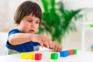
The Atkinson and Nardini (2008) model reveals that visuospatial ability begins around one month, when the child begins to have voluntary control of their eyes; at 3 months with the attempt to reach for objects; between 5 to 6 months with grasping objects; at 8 months with manual prehension; at 12 months with the manual exchange of objects; between 12 and 18 months they begin to build towers; between 3 and 4 years they create two-dimensional models and between 5 and 6 years they copy figures; furthermore by this age a right hemispheric dominance and asymmetry for these skills has already been established (Roselli, 2015; van der Ham & Ruotolo, 2017). Therefore, a delay in acquiring these developmental milestones could be a risk factor or warning sign of an alteration in visuospatial skills.
Neuropsychological rehabilitation of visuospatial skills
Neuropsychological rehabilitation is a procedure that aims to improve, as much as possible, the affected cognitive abilities of the patient in order to achieve optimal adaptation to their psychological, emotional, social, family and school/work life (Peña-Casanova et al., 1984). One of the goals of rehabilitating visuospatial skills is for the patient to be able to make copies and assemble objects with the greatest possible accuracy to their models; this implies not only having them copy but also stimulating preceding processes and providing strategies that allow better consolidation of visuospatial ability.
Some articles (Blázquez-Alisente et al., 2004; Serrano-Juárez et al., 2018) that have worked on the intervention of visuospatial skills have used activities that involve selective attention, eye movement, figure–ground, mental rotation, among others.
Activities for the neuropsychological intervention of visuospatial skills
Below are 6 activities that can be used for the neuropsychological intervention of visuospatial skills:
Visual scanning tasks
Eye movement is important for adequate scanning that allows detecting all components that make up a figure. The patient is asked to follow the tip of a pencil moving only their eyes; or on a computer a stimulus that moves randomly across the screen is created and the patient is asked to follow it only with their eyes.
Visuomotor coordination tasks
A task similar to the previous one is performed but in this case the patient is asked to follow it with their eyes and with the index finger of their dominant hand; later, they can be asked to do it with a pencil. Perform different paths with different shapes; straight and curved, and of different thickness, wide and thin. Create figures by joining dots.
Selective attention tasks
Carry out cancellation tasks teaching a scanning pattern from right to left and from top to bottom; in severe cases you can use their finger as a guide. Perform figure–ground tasks where they are asked to trace over with a different color all the figures they find.
Visual closure tasks
For a patient to begin to identify incomplete figures they must first identify them completely, so activities are done where they match complete objects and/or figures and progressively degrade them as they advance. Strategies such as completing the figure are used to train visual closure.
Spatial relations tasks
To improve the sense of laterality place a blue bracelet on the right hand and a red one on the left. Likewise, you can play “Simon says…” asking them to take steps forward, back, left or right. Draw a line down the middle of the page and ask them to place different objects above, below, to the left or to the right of it. Place three objects at different distances and with a different number of circles between each of them; then ask them to indicate which are closer and which are farther; they can be helped by the number of circles between each object.
Copying drawings
Ask the patient to make copies of figures but following a strategy taught by the therapist. For example, first start by identifying and copying the largest figures; then the medium ones and finally add the details; also each step can be done in a different color until copies similar to the model are achieved. Assembling and putting together puzzles.
Conclusions
Proper development of visuospatial skills is important for any individual since they have been associated with other processes such as calculation and writing; therefore, identifying and assessing these skills in time will allow the creation of programs, strategies and intervention activities that achieve early improvement, which could also impact other skills, processes and even adaptive behavior.
References
- Atkinson, J. (2002). The Developing Visual Brain. Oxford University Press. https://doi.org/10.1093/acprof:oso/9780198525998.001.0001
- Atkinson, J., & Braddick, O. (2011). Chapter 15—From genes to brain development to phenotypic behavior: “Dorsal-stream vulnerability” in relation to spatial cognition, attention, and planning of actions in Williams syndrome (WS) and other developmental disorders. En O. Braddick, J. Atkinson, & G. M. Innocenti (Eds.), Progress in Brain Research (Vol. 189, pp. 261–283). Elsevier. https://doi.org/10.1016/B978-0-444-53884-0.00029-4
- Atkinson, J., & Nardini, M. (2008). The neuropsychology of visuospatial and visuomotor development. Child neuropsychology: Concepts, theory and practice, 183–217.
- Blázquez-Alisente, J., Paúl-Lapedriza, N., & Muñoz-Céspedes, J. (2004). Attention and executive functioning in the neuropsychological rehabilitation of visuospatial processes. Rev Neurol, 38(5), 487–495.
- Doniger, G. M., Foxe, J. J., Murray, M. M., Higgins, B. A., & Javitt, D. C. (2002). Impaired Visual Object Recognition and Dorsal/Ventral Stream Interaction in Schizophrenia. Archives of General Psychiatry, 59(11), 1011. https://doi.org/10.1001/archpsyc.59.11.1011
- D’Souza, D., Booth, R., Connolly, M., Happé, F., & Karmiloff-Smith, A. (2016). Rethinking the concepts of ‘local or global processors’: Evidence from Williams syndrome, Down syndrome, and Autism Spectrum Disorders. Developmental Science, 19(3), 452–468. https://doi.org/10.1111/desc.12312
- Kravitz, D. J., Saleem, K. S., Baker, C. I., & Mishkin, M. (2011). A new neural framework for visuospatial processing. Nature Reviews Neuroscience, 12(4), 217–230. https://doi.org/10.1038/nrn3008
More references on the rehabilitation of visuospatial skills
- Labos, E., Slachevsky, A., Fuentes, P., & Manes, F. (2008). Treatise of Clinical Neuropsychology. Buenos Aires: Akadia.
- Peña-Casanova, J., Pamies, M. P., García, J. S., & Pulido, J. H. (1984). Rehabilitation of Aphasia and Associated Disorders. Masson.
- Roselli, M. (2015). Neuropsychological development of visuospatial and visuoconstructive skills. Revista Neuropsicología, Neuropsiquiatría y Neurociencias, 15(1), 175–200.
- Serrano-Juárez, C. A., Prieto-Corona, D. M. B., & Yáñez-Téllez, M. G. (2018). Neuropsychological intervention in a case of a girl with Williams Syndrome. Cuadernos de Neuropsicología/Panamerican Journal of Neuropsychology, 12(2).
- Stiles, J., Akshoomoff, N. A., & Haist, F. (2020). Chapter 17—The development of visuospatial processing. En J. Rubenstein, P. Rakic, B. Chen, & K. Y. Kwan (Eds.), Neural Circuit and Cognitive Development (Second Edition) (pp. 359–393). Academic Press. https://doi.org/10.1016/B978-0-12-814411-4.00017-2
- van der Ham, I. J. M., & Ruotolo, F. (2017). On inter- and intrahemispheric differences in visuospatial perception. En Neuropsychology of space: Spatial functions of the human brain. (pp. 35–76). Elsevier Academic Press. https://doi.org/10.1016/B978-0-12-801638-1.00002-1
If you liked this post about the rehabilitation of visuospatial skills, you might be interested in these publications from NeuronUP:


“This article has been translated. Link to the original article in Spanish:”
Rehabilitación neuropsicológica de las habilidades visoespaciales
 Occupational Therapy in Patients with Acquired Brain Injury


Leave a Reply