PhD in Clinical Health Psychology María J. García-Rubio, together with specialist in clinical neuropsychology and major neurocognitive disorder Nancy Navarro, explain in this article what a neurological examination is, as well as its techniques and clinical applicability.
Introduction
The knowledge of the structure and function of the nervous system (NS) requires the combination of certain disciplines such as physiology, medicine, psychology, and biology, among others. Therefore, many of these compose neuroscience and the milestones that accompany it as a specific branch for the study of the NS in its healthy and pathological versions.
To carry out the scientific studies that collect information about the development of the NS, its parts and mode of operation, a common methodology is required among these disciplines that come together around the neurological examination. And from there arise the neurological examination techniques.
What is the neurological examination?
The neurological examination is the action through which one can draw precise conclusions about the structure and function of various aspects of the nervous system (González and López, 2013), with a key factor being the type of neurological examination technique and the mechanism underlying it.
Neurological examination techniques
The development of neurological examination techniques dates back to the evolution of the concept of the brain, which coincides with the time of some well-known philosophical theories such as that of Hippocrates (c. 460–370 BCE), who described the brain as the seat of experience and intelligence.
Andreas Vesalius (1514–1564) was the first to describe the anatomy of the brain through illustrations gathered in his work “De humani corporis”; and René Descartes (1596–1650) describes the brain as a complex machine, similar to the heart, that controls the complex actions of the human being.
Pneumoencephalography
In 1919, Walter Dandy (1886–1946) developed pneumoencephalography, a method that consisted of taking an X-ray of the brain by replacing the cerebral fluid with air, oxygen, or helium.
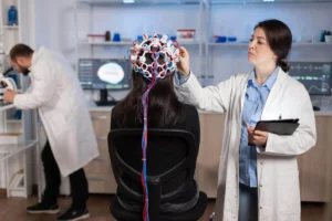
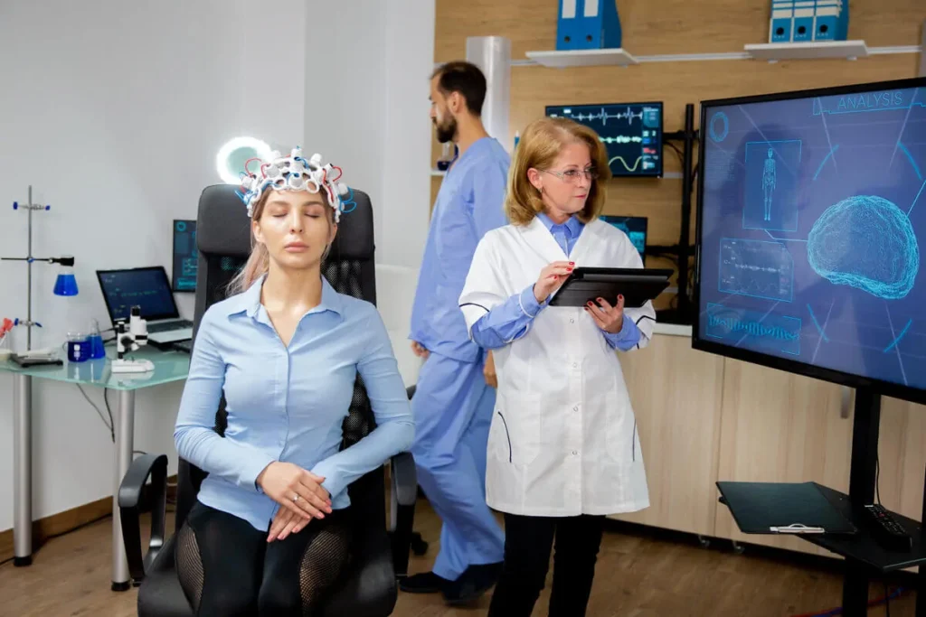
The electroencephalogram
After this first neurological examination technique came the electroencephalogram from the German Hans Berger (1873–1941) as a method capable of revolutionizing neurological assessment known since then. With EEG, the researcher does not have an X-ray of the brain, but can know the timings and brain areas in which neural activity occurs based on postsynaptic potentials recorded at the person’s scalp.
Other neurological examination techniques and their variants
Subsequently, other neurological examination techniques included angiography originating from lobotomy practices in the 20th century. However, it was at the end of the 1960s when functional neurological examination techniques emerged such as magnetoencephalography (MEG) by Cohen (1968), computed tomography (CT), and magnetic resonance imaging (MRI), along with the development of positron emission tomography (PET) (Posner and Raichle, 1994).
Finally, in the 1980s and thereafter, new variants of these same neurological examination techniques have been developed, enabling researchers to extract increasingly precise and informative brain data.
Examples include transcranial magnetic stimulation (EMT/TMS), functional magnetic resonance imaging (fMRI) and its various applications, among which BOLD signals, T1 and T2 stand out. Also, since 1994 there has been access to neurological examination from imaging data obtained with more current techniques such as diffusion tensor imaging that allows visualization of the trajectory of nerve fibers, among others (Fuller, 2020).
Neurological examination methods
Throughout this post, we will provide a brief summary of the neurological examination methods that exist and we will focus especially on the functional methods that are most used today due to their major contribution in cognitive neuroscience studies with human subjects.
Lesion methods
Lesion methods are neurological examination techniques that use brain lesions as hypotheses to understand brain performance related to a behavior, that is, they analyze damaged brain structures and their influence on behavior.
Their methodology is broad for the study of different scenarios, both for lesions that have occurred spontaneously due to acquired brain injury, as well as lesions resulting from a surgical procedure (Humphreys et al., 2021).
Among these methods are macroscopic and microscopic analysis, whose purpose is to provide information on the morphological and architectural composition of nervous tissue post-mortem. This is carried out through procedures such as fixation, staining, or sectioning, to determine chemical and molecular nature at the macro- or microscopic level.
Instrumental methods
These methods allow neurological examination through the recording and observation of brain performance during different tasks, using instruments specifically adapted to control variables that indirectly affect brain functioning (Marinescu et al., 2018).
Some neurological examination techniques that fall within this category are sensory techniques, derived from the sensory division paradigm, according to which different input information is manipulated so that several controlled stimuli reach both cerebral hemispheres at once and compete for arrival. Notable among these are dichotic hearing, separate visual fields, and dihapic palpation (Maes et al., 2017; Vendetti et al., 2015).
Motor techniques are also instrumental neurological examination methods, such as motor interference, which requires the participant to perform two motor tasks simultaneously, for example, the tapping test.
Functional methods
Functional methods are those that record changes in brain activities by manipulating behavioral variables. Within this category, electromagnetic and metabolic neurological examination techniques stand out.
Electromagnetic techniques: among them are electroencephalography, evoked potentials and magnetoencephalography.
— Electroencephalogram
It is a non-invasive technique that allows study of the central nervous system. From this, one can access brain examination while different tasks are performed in response to distinct stimuli.
The method allows amplification of the brain bioelectrical activity by detecting the activity of electric fields generated by postsynaptic potentials of pyramidal neurons in the cortex. The gamma, beta, theta, and delta wave patterns generated during the process are associated with behavior and consciousness, these being measured by their frequency and amplitude.
The main clinical conditions that can be detected by this technique include: diagnosis for epileptic disorders, classification of sleep pathologies, and alterations in level of consciousness (Keren et al., 2018).
— Evoked potentials (EP)
They measure changes in brain electrical activity through an external sensory stimulus. The activity is detected by electrodes placed on the scalp, providing information as an indicator of neural activity (Bestmann and Krakauer, 2015).
These potentials are used for the assessment of the auditory pathway and in studies of: newborns at risk of hearing loss, degenerative and demyelinating diseases (Norcia et al., 2015).
— Magnetoencephalography
It consists of a test that measures the magnetic fields generated by the electrical currents of the brain and detects changes in specific areas. The procedure consists of one or more sensors that move over the surface of the skull and produce an isocontour map representing different intensities of the magnetic field.
In clinical practice this neurological examination technique is used to locate the focus of epileptic seizures (Förster et al., 2020).
Metabolic techniques: those neurological examination instruments that allow detection of the level of cerebral metabolic activity. For this purpose, the following techniques are used: positron emission tomography, single-photon emission computed tomography, and functional magnetic resonance imaging.
— Tomography by positron emission (PET)
It is used to explore neuronal metabolic activity using radiotracers that allow measurement of blood flow, dopamine transporter mechanisms or serotonin reuptake.
This process does not pose risks to the patient; on the contrary, the radioactive molecules emit particles called positrons to determine the brain region to be explored and create a color image of the brain (O’Neill et al., 2015). This method is widely used in the detection of neurodegenerative diseases.
— Single-photon emission computed tomography (SPECT)
It is a technique used in the field of nuclear medicine. It combines two-dimensional images that, when combined, form a single three-dimensional image.
This neurological examination technique is widely used in the evaluation of cerebrovascular disease, and in patients with suspected dementia syndrome and/or traumatic brain injury (Pelegrí Martínez, et al., 2017).
— Magnetic Resonance (fMRI)
It allows measurement of regional metabolism by detecting oxygen in the blood vessels of the brain. The physiological explanation shows that as neurons become more active, they use greater amounts of oxygen, which produces a reduction of oxygen in the blood (Keren et al., 2018).
Neurons send signals to blood vessels to generate vasodilation and thus transport oxygen. fMRI aligns this process with great precision in the localization within brain structures.
The use of these neurological examination techniques is diverse; currently, they are used in the field of Cognitive and Affective Neuroscience with healthy populations or with neuropathological diagnoses (Atenas et al., 2018).
Conclusion
This blog post has introduced the development and state of neurological examination techniques, making a special emphasis on functional techniques from which imaging data are extracted, especially related to human cognitive function at rest and/or while performing a task with high or low demand, for example.
Likewise, these techniques allow the neurological examination of both the healthy and the pathological brain.
In conclusion, neurological examination techniques constitute one of the most current, innovative, and developing methods in neuroscience at the national and international level, due to their applicability as a procedure and the quality and suitability of their results.
Bibliography
Atenas, T. L., Díaz, E. C., Quiroga, J. C., Arancibia, S. U., & Rodríguez, C. C. (2018). Functional magnetic resonance imaging: Basic principles and application in the neurosciences. Radiología, 60(5), 368-377. https://doi.org/10.1016/j.rxeng.2018.04.001
Bestmann, S., & Krakauer, J. W. (2015). The uses and interpretations of the motor-evoked potential for understanding behaviour. Experimental brain research, 233, 679-689. https://doi.org/10.1007/s00221-014-4183-7
Förster, J., Koivisto, M., & Revonsuo, A. (2020). ERP and MEG correlates of visual consciousness: The second decade. Consciousness and cognition, 80, 102917. https://doi.org/10.1016/j.concog.2020.102917
Fuller, G. (2020). Easy Neurological Examination. Elsevier Health Sciences.
González, N. C., & López, J. A. T. (2013). Manual for the neurological examination and higher brain functions. Manual Moderno.
Humphreys, C. A., Smith, C., & Wardlaw, J. M. (2021). Correlations in post‐mortem imaging‐histopathology studies of sporadic human cerebral small vessel disease: a systematic review. Neuropathology and applied neurobiology, 47(7), 910-930. https://doi.org/10.1111/nan.12737
Keren, H., O’Callaghan, G., Vidal-Ribas, P., Buzzell, G. A., Brotman, M. A., Leibenluft, E., … & Stringaris, A. (2018). Reward processing in depression: a conceptual and meta-analytic review across fMRI and EEG studies. American Journal of Psychiatry, 175(11), 1111-1120. https://doi.org/10.1176/appi.ajp.2018.17101124
Maes, C., Gooijers, J., de Xivry, J. J. O., Swinnen, S. P., & Boisgontier, M. P. (2017). Two hands, one brain, and aging. Neuroscience & Biobehavioral Reviews, 75, 234-256. https://doi.org/10.1016/j.neubiorev.2017.01.052
Marinescu, I. E., Lawlor, P. N., & Kording, K. P. (2018). Quasi-experimental causality in neuroscience and behavioural research. Nature human behaviour, 2(12), 891-898. https://doi.org/10.1038/s41562-018-0466-5
Norcia, A. M., Appelbaum, L. G., Ales, J. M., Cottereau, B. R., & Rossion, B. (2015). The steady-state visual evoked potential in vision research: A review. Journal of vision, 15(6), 4-4. https://doi.org/10.1167/15.6.4
O’Neill, G. C., Barratt, E. L., Hunt, B. A., Tewarie, P. K., & Brookes, M. J. (2015). Measuring electrophysiological connectivity by power envelope correlation: a technical review on MEG methods. Physics in Medicine & Biology, 60(21), R271. 10.1088/0031-9155/60/21/R271
Pelegrí Martínez, L., Kohan, A. A., & Vercher Conejero, J. L. (2017). Optimization of protocols and the use of contrast agents in computed tomography for positron emission tomography equipment. Radiología, 64-74.
Posner, M.I., & Raichle, M.E. (1994). Images of Mind. Scientific American Library.
Vendetti, M. S., Johnson, E. L., Lemos, C. J., & Bunge, S. A. (2015). Hemispheric differences in relational reasoning: novel insights based on an old technique. Frontiers in human neuroscience, 9, 55. https://doi.org/10.3389/fnhum.2015.00055
If you enjoyed this article about neurological examination techniques, you may also be interested in the following articles:


“This article has been translated. Link to the original article in Spanish:”
Técnicas de exploración neurológica: conceptos básicos y aplicabilidad clínica
 Cognitive Intervention Program for People with Williams Syndrome


Leave a Reply