
Neuropsychologist Javier Esteban Libiano lays out in this article all the details about how our brain encodes smells.
Smell is, together with taste, a chemical sense. The stimuli received by both senses interact chemically with their receptors.
Smell helps identify foods and avoid those that are spoiled or not suitable for consumption. It helps many species track prey or detect predators, as well as identify friends, enemies, and receptive mates.
For humans, smells have the peculiar ability to evoke memories. Smell activates brain regions related to emotion, learning and memory.
Could we use this information to exert a neurorrehabilitative effect on different cognitive abilities in subjects with some type of brain injury or cognitive impairment associated with various pathologies?
The stimulus, nature and characteristics
There is increasing evidence every day that sensory stimulation has an effect on the maintenance and improvement of cognitive abilities -such as perception, language, praxis, gnosias, attention, memory, executive functions, orientation, reasoning and motivation,- in people with processes of cognitive decline, cerebrovascular accidents (strokes) or other factors that have caused brain damage.
The olfactory stimulus, technically known in English as odorants, consists of volatile substances that have a molecular weight between 15 and 300 g/mol (grams per mole). Almost all odorous compounds are lipid-soluble and of organic origin, although many substances that meet these criteria have no smell.
Odoriferous substances must be water-soluble to be able to dissolve in the superficial mucous layer and reach the olfactory cilia, on whose membranes they bind to specific receptors. At sufficiently high concentrations they produce a depolarization of the cell membrane that is transmitted along the axon as an action potential.
It is believed that in smell there would be some basic qualities that are registered by specific receptors.
Since substances that belong to the same olfactory group have similar molecular sizes, it seems possible that the membrane of an olfactory cilium, projections of the receptor cells that penetrate the mucosa and receive the stimulus, react only to a certain molecular size.
New research suggests that a sensory cell expresses only one type of receptor.
A study from the University of California found that some odors could increase people’s cognitive capacity by conducting an experiment in which a sample of individuals was exposed to the scent of a fragrance while they slept. This research opens the door to non-invasive neurorrehabilitative treatments to fight neurodegenerative diseases or various brain injuries.
The receptors
We have six million olfactory receptor cells (bipolar neurons), which are located within two portions of the mucous membrane (the olfactory epithelium).
In humans, the olfactory epithelium covers a small area of both nasal cavities, being located in the upper part of the nasal cavity, on the edge of the superior turbinates and on the opposite surface of the nasal septum.
The receptors for smell are found in the sensory epithelium, which is made up of sustentacular or supporting cells and sensory cells or olfactory receptor cells.
Olfactory receptor cells are bipolar neurons, whose cell bodies are in the olfactory mucosa that covers the cribriform plate, an opening at the base in the rostral part of the brain.
Between the olfactory receptor cells there are supporting cells, which contain enzymes that destroy odorous molecules and thus help prevent the olfactory receptors from being altered.
The olfactory region also contains numerous small mucous glands, the Bowman glands, whose secretion forms a thin terminal film that coats the olfactory mucosa.
The distal segment of the sensory cell tapers to form a thin stalk that slightly protrudes above the surface of the olfactory epithelium. This olfactory protrusion is covered by numerous olfactory cilia. Proximally, the oval cell body continues with a thin process that, along with the other processes, is wrapped by Schwann cells.
Stimulus-receptor interaction
Researchers acknowledge that the olfactory cilia contain molecular receptors that are stimulated by odorous molecules.
Jones and Reed identified a particular G protein, which they called Golf. This protein can activate an enzyme that catalyzes the synthesis of cyclic adenosine monophosphate (cyclic AMP, a nucleotide that acts as a second messenger in different biological processes), which in turn can open sodium channels and depolarize the membrane of the olfactory cell.
G proteins serve as a link between metabotropic receptors and ion channels: when a ligand binds to a metabotropic receptor (signal transduction mechanisms, often G proteins, to activate a series of intracellular events using second messenger chemicals), the G protein opens any ion channel directly or indirectly, by activating the production of a second messenger. The discovery of Golf suggested that the olfactory cilia contained odor receptors coupled to this G protein.
Buck and Axel discovered a family of genes that encodes a family of olfactory receptor proteins. In humans there appear to be between 500 and 1000 different receptors, each sensitive to a different odor. Odorous molecules bind to these receptors and the coupled G proteins cause the opening of sodium channels, producing depolarizing action potentials.
The olfactory cognitive stimulation opens a field of intervention in the realm of neuroplasticity, known as the brain’s capacity to recover, restructure, recombine, remodel, reform, reorganize and adapt to new situations.

Subscribe
to our
Newsletter
The olfactory nerves
Olfactory receptor cells send through the surface of the mucosa a process that divides into 10 or 20 cilia, which penetrate the mucus layer.
These processes gather to form the olfactory nerves that pass through the openings of the cribriform plate to reach the olfactory bulb. Approximately thirty-five bundles of axons, surrounded by glial cells, pass through the bone via small openings of the cribriform plate.
The processes terminate in the olfactory glomeruli of the olfactory bulb, where they form synapses with the dendrites of mitral cells.
Two other paired nerves accompany the olfactory nerve from the nasal cavity to the brain: the terminal nerve and the vomeronasal nerve.
The terminal nerve is formed by a fascicle of thin nerve fibers that run from the nasal septum through the cribriform plate to the lamina terminalis, entering the brain below the anterior commissure. This fascicle contains numerous nerve cells and is considered an autonomic nerve.
The vomeronasal nerve that runs from the vomeronasal organ to the accessory olfactory bulb is developed in lower vertebrates and in humans is only demonstrable during embryonic development.
The olfactory mucosa also contains free nerve endings of the axons of the trigeminal nerve. These nerve endings mediate pain sensations that can be produced by sniffing some chemical irritants such as ammonia.
The olfactory bulbs
The olfactory bulbs are thickened regions at the end of the olfactory tract, which receive afferents from the olfactory receptors. They are located at the base of the brain, at the end of the elongated olfactory tracts.
Each receptor cell sends a single axon to the olfactory bulb, where it synapses with the dendrites of mitral cells. These synapses take place in the axonal complex and in the dendritic arborizations called olfactory glomeruli.
There are approximately 10,000 glomeruli, each of which receives afferents from a bundle of approximately 2,000 axons.
Neural pathways
Path of the stimulus
In summary, the olfactory receptors consist of bipolar neurons located in the olfactory epithelium, which lines the roof of the nasal sinuses, in the bone that lies beneath the frontal lobes.
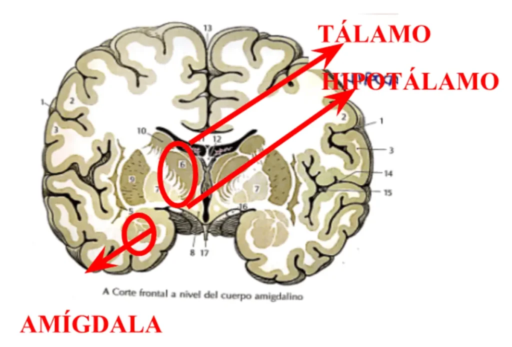
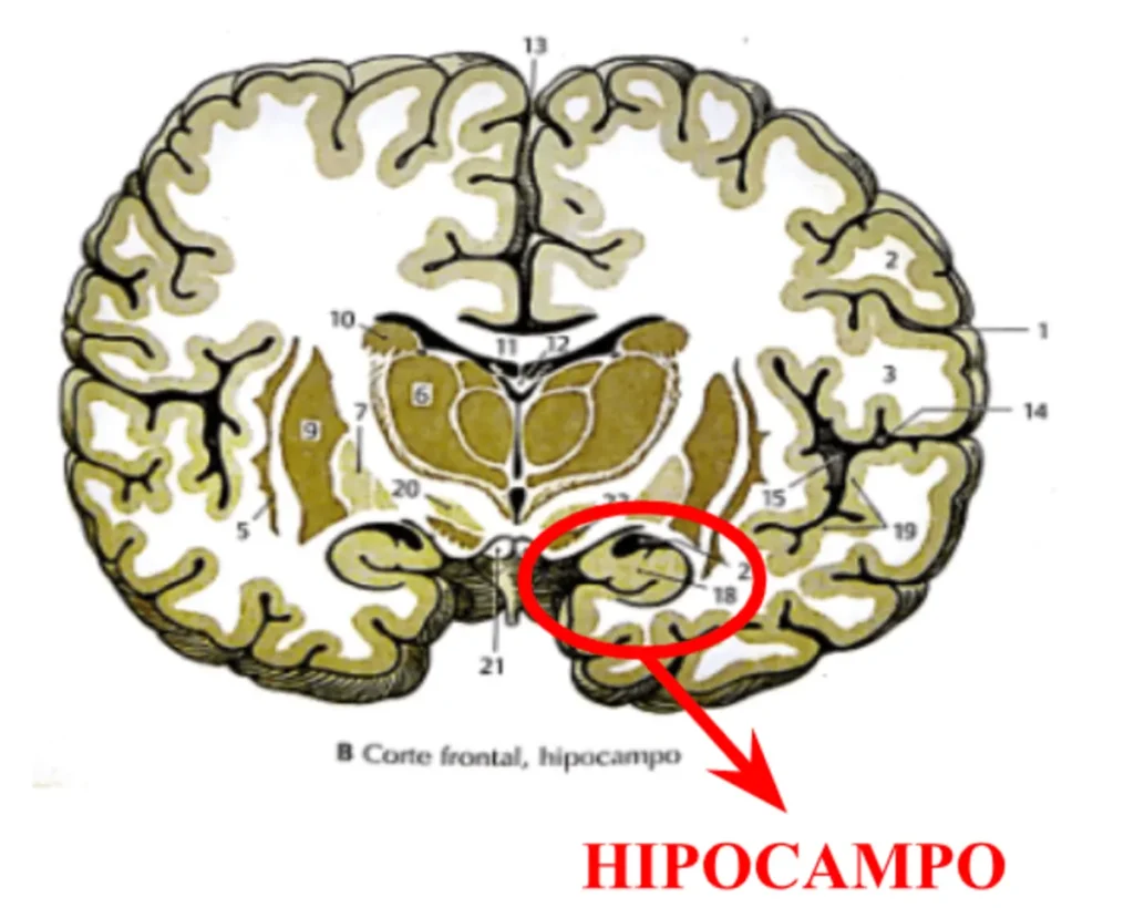
The receptors send endings across the surface of the mucosa, which divide into cilia. The membranes of these cilia contain receptors that detect odorous molecules dissolved in the air that reach the olfactory mucosa. The axons of the olfactory receptors pass through the openings of the cribriform plate to the olfactory bulbs, where they form synapses in the glomerulus with the dendrites of mitral cells. These neurons send axons, through the olfactory tracts, to the brain, mainly to the amygdala (related to emotions), the piriform cortex (information processing) and the entorhinal cortex (memory, orientation and learning). The hippocampus (learning and memory), the hypothalamus and the orbitofrontal cortex (decision-making), receive olfactory information indirectly.
Vomeronasal organ
Most mammals have another organ that responds to environmental chemicals: the vomeronasal organ, which is a sensory epithelium located inside a mucous sac of the nasal septum, important in reptiles for tracking food. It also plays an important role in the animal response to pheromones (chemical substances produced by animals that affect reproductive physiology and behavior).
Different regions involved

Axons of the olfactory tract project directly to the amygdala and to two regions of the limbic cortex: the piriform cortex and the entorhinal cortex.
- The amygdala, related to emotion, sends olfactory information to the hypothalamus, which is related to food intake and emotion.
- The entorhinal cortex, related to learning and memory, sends olfactory information to the hippocampus, which is also related to learning and memory.
- The piriform cortex, related to memory and learning, sends olfactory information to the hypothalamus and to the orbitofrontal cortex, related to memory and emotion, via the dorsomedial nucleus of the thalamus, related to memory and learning.
- The orbitofrontal cortex, in addition to olfactory information, also receives gustatory information. Therefore, it could be involved in combining taste and smell in flavors.
- The hypothalamus receives a considerable amount of olfactory information, which is likely important for the acceptance or rejection of foods and for the olfactory control of reproductive processes.
We thus see that through cognitive stimulation, using smell and the different odors we can encounter as a tool, we will activate brain regions related to memory, learning, emotion, decision-making, information processing… In this way, an activity as simple as recognizing different odor stimuli, can result in the improvement or slowing down of different processes of cognitive decline, offering us a field of intervention that can at the same time be combined with other cognitive stimulation techniques.
Perception of specific odors
People can recognize more than 10,000 different odors. Even if we had several hundred different olfactory receptors, or even a thousand, many odors would remain unexplained.
How can we use a relatively small number of receptors to detect so many different odors?
Recognition of a particular odor is a matter of recognizing a particular configuration of activity in the glomeruli. The task of chemical recognition becomes a task of spatial recognition.
The spatial patterns of olfactotopic information are maintained in the olfactory cortex. Probably, the brain recognizes particular odors by recognizing the different patterns of activation that occur.
Although most odors are produced by mixtures of many different chemical substances, we identify them as belonging to a specific object, for example, the smell of coffee or the smell of cigarette smoke.
Bibliography
- N. Carlson. (2006). Physiology of Behavior. Pearson Publishing, Madrid
- W. Kahle. (2003). Atlas of Anatomy. Volume 3. Nervous system and sensory organs. Omega Publishing, Barcelona
- Woo, C; Miranda, B; Sathishkumar, M; Dehkordi-Vakil, F; Yassa, M and Leon, M. (2023). Overnight olfactory enrichment using an odorant diffuser improves memory and modifies the uncinate fasciculus in older adults. Frontiers in Neuroscience.
If you liked this blog post about the neuroscience of smell in neurorehabilitation, you will likely be interested in these NeuronUP articles:


“This article has been translated. Link to the original article in Spanish:”
Neurociencia del olfato en neurorrehabilitación: ¿Cómo codifica el cerebro los olores?
 What will I find in NeuronUP from now on?


Leave a Reply